Journal of
eISSN: 2574-9943

Cutaneous endometriosis (CEM) is a rare cutaneous manifestation of a very common gynecologic disease among middle-aged women. Cutaneous lesions are commonly seen at sites of previous surgical scars, but can also arise de-novo in normal epithelium. The clinical presentation alone can vary widely and mimic other common dermatologic conditions. Histopathologic examination is often needed to confirm the diagnosis. Here we will present both a primary and secondary case of CEM in attempts to portray the wide variation in physical exam findings so CEM may be considered in the differential for cutaneous masses in the umbilicus.
Keywords: Cutaneous endometriosis; Women; Dermatologic conditions; Endometrial glands; Laparoscopy
Endometriosis is a common condition among women that is characterized by the presence of endometrial tissue outside of the uterine cavity. This tissue has been reported in a wide variety of locations throughout the body, with a rare number of cutaneous cases documented. Cutaneous involvement accounts for 0.5-1% of endometriosis cases and is most commonly located at sites of previous surgical scars (secondary CEM). Primary (spontaneous) CEM accounts for 30% of cutaneous cases. Both have a predilection for the umbilical region. Lesions may develop swelling, pain, and bleeding that parallels the timing of the menstrual cycle, which may clue the physician into the diagnosis. We present two cases of CEM with different clinical presentations and physical examination findings that share the same pathogenesis confirmed by histopathologic examination.
Case 1: Secondary Cutaneous Endometriosis: A 30 year-old African-American female presented in the clinic with complaints of a slowly enlarging umbilical mass that had been present for one year. The lesion was painful and bled on occasion, mostly around her menstrual cycle. Skin examination revealed a 2.5cm hyper pigmented, pedunculated nodule inferior to a well-healed surgical scar from a previous laparoscopic tubal ligation (Figure 1). The patient denied any pelvic symptoms and was never diagnosed with endometriosis or had difficulty conceiving. Histopathological examination revealed endometrial glands, stroma, and hemorrhage within the dermis (Figure 2). No evidence of malignancy was noted. Patient was initiated on oral contraceptives with a plan to perform surgical excision in the near future.

Figure 1: Hyperpigmented, pedunculated umbilical nodule measuring 2.5 cm in a 30-year-old African-American female inferior to a well-healed surgical scar.
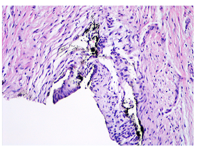
Figure 2: Microscopic examination of the umbilical nodule shows a glandular structure lined by low columnar epithelium. The surrounding stroma consists of spindle cells, H&E.
Case 2: Primary Cutaneous Endometriosis: A 30 year-old Caucasian female presented in the clinic with complaints of two umbilical lesions that presented one year prior. She reported intermittent pain, swelling, and bleeding of the lesions in association with her menstrual cycles. The patient denied a history of any abdominal or pelvic surgical procedures or trauma in the area of the lesions. However, she did report dysmenorrhea. Skin examination revealed two 2-3 mm hyper pigmented papules within the umbilicus (Figure 3). Histopathologic examination showed low cuboidal-lined glandular spaces in the dermis. The glandular cells were positive for EMA and AE1/3. A CD10 stain was positive in the immediately surrounding stroma, and iron stain was focally positive. Similar to Case 1, oral contraceptives were prescribed and surgical excision was recommended by OB/Gyn.
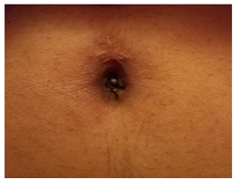
Figure 3: A 30-year old Caucasian female with two 2.5 mm hyperpigmented papules in the umbilicus that have been present for one year .The lesions were associated with cyclical pain, swelling, and bleeding associated with her menstrual cycle.
Endometriosis, or the ectopic implantation of endometrial tissue outside of the uterus, is an extremely common condition affecting up to 6-15% of women of reproductive age [1]. The endometrial tissue is responsive to hormones that affect the menstrual cycle and presents in middle-aged women as cyclical pelvic pain, dysmenorrhea, dyspareunia, infertility, and occasionally pain on defecation. The most common sites of implantation include the pelvic peritoneum, ovaries, and rectovaginal septum. Cutaneous endometriosis is a rare phenomenon that comprises only 1-5% of all cases and is most commonly found in the umbilicus, also known as Villar’s nodule. Umbilical endometriosis is most commonly seen in reproductive age women (mean age 36.3 years) who present with a red-brown nodule accompanied by cyclical pain at the site. Subcutaneous endometriomas may be present, and they appear as red, blue, black, or flesh colored nodules on the abdomen. Cyclical bleeding, pain, swelling, and abdominal discomfort are all common symptoms associated with CEM. A clinicopathological review of 34 cases concluded that pain was the most common presentation for extragenital endometriosis, but was present in only 41.2% of patients [2]. The lesions can be a diagnostic challenge due to the rarity of this diagnosis and heterogenous clinical presentation. The definitive diagnosis relies on histopathologic confirmation of tissue containing endometrial stroma and glands usually from a biopsy.
Cutaneous endometriosis can be primary or secondary; secondary is most often seen at sites of prior surgical incision, usually a gynecologic procedure such as cesarean section, laparoscopy, hysterectomy, or Bartholin gland cyst removal [3]. Pathogenesis of secondary cutaneous endometriosis can often be attributed to iatrogenic dissemination, as incision sites provide routes of seeding from internal primary sources. The definite pathogenesis for primary cutaneous endometriosis is not well understood, but multiple theories exist to explain the migration of uterine tissue to ectopic sites including retrograde migration through fallopian tubes, coelemic metaplasia, direct spread, and lymphatic or hematogenous spread [4]. For patients with preexisting pelvic endometriosis, lymphatic or hematogenous spread is the most probable etiology of ectopic uterine tissue. It is possible that the umbilicus represents a physiological scar and may have an innate predilection for endometrial tissue [1]. Another theory suggests metaplasia of urachal remnants as the cause of cutaneous endometriosis [4].
The variation in presentation reveals a diagnostic challenge for an umbilical nodular lesion in a young female. Our patients share similar age and demographical characteristics, but vary in other aspects of clinical presentation. First, one patient has a significant surgical history contributing to the nodular formation, while the other patient reports spontaneous onset with no trauma to the area. The cyclical symptoms of pain and bleeding were similar and both paralleled the menstrual cycle. The physical examination is strikingly different between the two case reports, which is why CEM always must remain on the differential for umbilical lesions. Both diagnoses were supported by histopathologic evidence of ectopic endometrial tissue. The differential diagnosis includes benign lesions such as keloid, pyogenic granuloma, umbilical hernia, melanoma, suture granuloma, and omphalith or foreign body. Umbilical malignancy, also known as Sister Mary Joseph’s nodule, must also be considered and ruled out. Table 1 highlights distinguishing features to assist in the clinical diagnosis.
Condition |
Characteristics |
Cutaneous endometriosis |
Nodule that presents with pain, Pruritis, odor, swelling, and bleeding associated with the menstrual cycle |
Nodular melanoma |
Uniform dark black/brown smooth nodule; ulceration and bleeding are late complications. |
Usually presents as a relatively small lesion with average thickness of 2 mm |
|
Sister Mary Joseph’s nodule |
Firm, Indurated plaques or nodules, usually with vascular appearance; can be fissured or ulcerated with discharge. |
Associated symptoms: anorexia, weight loss, and/or epigastric pain. |
|
Past medical history of primary malignancy increases risk |
|
Pyogenic granuloma |
Red or reddish-brown pedunculated, friable papule with color of hyperkeratosis at the base +/- crust and erosion; bleeds easily. |
Usually occurs in areas of trauma or around the mouth in pregnant females |
|
Keloid |
Flesh-colored, pink-red, or hyperpigmented, smooth, and firm papule +/- pain and pruritus. |
Grow gradually with claw-like extensions beyond wound site. |
|
Can be constantly pruritic or painful, but symptoms usually constant |
|
Umbilical hernia |
Flesh-colored, soft, reducible nodule. |
Worsened with increased intra-abdominal pressure (lifting, standing, straining, coughing) |
|
Suture granuloma |
Pustule or erythematous nodules within a suture site following surgery |
Omphalith |
Firm, gray to dark brown compact papule or nodule within the umbilicus. |
Poor personal hygiene leading to sloughed epithelial cells and sebum accumulation |
Table 1: The differential diagnosis of an umbilical nodule.
The literature estimates a delay in diagnosis for CEM with an average time to diagnosis of 17.8 months [1]. Fine needle aspiration (FNA) can be utilized as an alternate diagnostic modality in lesions with high pretest probability for endometriosis. Advantages to FNA include: less invasive than biopsy, accurate, and quick. Cytology smears characteristic for endometriosis show epithelial cellular and stromal components admixed with hemorrhage and hemosiderin laden macrophages on a background of inflammatory cells [5]. Hormone levels alter cell structure and composition, so findings change between the proliferative and secretory phase of the menstrual cycle. Utilization of FNA for suspicious lesions may improve clinical diagnostic accuracy, decrease long-term manifestations of endometriosis, and optimize quality of life for these patients.
There are limited treatments for CEM with surgical excision the only definitive treatment option. Surgery is often performed at the end of the menstrual cycle when the ectopic tissue is the smallest and least active due to hormonal variations. Surgical technique revolves around the size and depth of the lesion and must be individualized for each patient. Gonadotropin-releasing hormones (GnRH), danazol, and oral contraceptive pills may be used to decrease the size of cutaneous lesions pre-operatively via hormonal control. It is recommended that all patients be evaluated by a gynecologist for coexisting internal endometriosis, which is found in around 15% of patients [6]. The prognosis of umbilical CEM is good, and the recurrence rate is extremely low if complete excision is achieved. The risk of scar tissue transformation from complete excision has been noted in the literature, so follow-up may be warranted [6].
In conclusion, physicians must have a high level of suspicion for CEM in patients presenting with symptomatic and asymptomatic umbilical nodules. A clinical evaluation with supporting histopathologic analysis confirms the diagnosis and allows prompt surgical excision of the lesion. This not only provides symptomatic relief, but also probable cure if clear margins are obtained in the procedure.
Cutaneous endometriosis is a rare cutaneous manifestation of a very common gynecologic disease in middle-aged women. Cutaneous manifestations widely vary in clinical presentation, and may be associated with bleeding, swelling, or intermittent pelvic or abdominal pain that parallels timing of the menstrual cycle. The majority of CEM cases are found in previous surgical incision sites, but de-novo lesions arising in normal epithelium comprise around 30% of cases. Cutaneous endometriosis has a predilection for the umbilicus and should be included in the differential for nodular umbilical lesions. Histopathologic confirmation depicting endometrial glands and stroma is required for definitive diagnosis, but FNA may be utilized as a diagnostic modality in patients with high pretest probability. Surgical excision is the only definitive treatment, but medications can be used to shrink the lesion pre-operatively through hormonal control. All patients should be evaluated by a gynecologist for the presence and management of internal endometriosis. A high level of suspicion can shorten the time to diagnosis, decrease severity of long-term effects of the disease, and improve patient quality of life.
We would like to acknowledge Dr. Pam Martin for her contribution and support. No additional funding was required.
There are no conflicts of interest pertinent to our case series.

© . This is an open access article distributed under the terms of the, which permits unrestricted use, distribution, and build upon your work non-commercially.