Journal of
eISSN: 2373-4396


Case Report Volume 2 Issue 1
1Department of Cardiology, CHU de Poitiers, France
2Department of Pathology and Laboratory Medicine, CHU de Poitiers, France
Correspondence: Rodrigue Garcia, CHU de Poitiers, Department of Cardiology, F-86021 Poitiers, France, Tel 33603847546, Fax 33549444010
Received: December 30, 2014 | Published: January 6, 2015
Citation: Garcia R, Taris M, Varroud-Vial N, Christiaens L (2015) Unusual Cause of Stem I: An Intramyocardial Metastasis. J Cardiol Curr Res 2(1): 00044. DOI: 10.15406/jccr.2015.02.00044
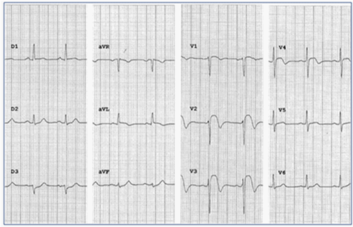
A 66-year-old patient was admitted to the emergency unit for chest pain. The electrocardiogram showed a ST-segment elevation in leads V2-V4 and a slight ST-segment depression in lead III. The coronary angiography was normal but CMR imaging and CT scan revealed a tumor surrounding the left anterior artery. Finally, further investigations disclosed a metastasis of squamous cell carcinoma lung cancer.
Keywords: heart neoplasms, acute coronary syndrome
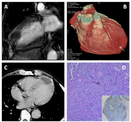
A 66-year-old patient, with a medical history of non-Hodgkin lymphoma in remission for 5years, was admitted to the cardiac emergency unit for chest pain. The electrocardiogram showed a ST-segment elevation in leads V2-V4 and a slight ST-segment depression in lead III (Figure 1). The troponin count was 10 fold higher than the upper limit but the coronary angiography immediately carried out was normal. In echocardiography, the apex was akinetic. A cardiac magnetic resonance imaging was performed and showed one intramyocardial mass of 30 x 42 x 38 mm at the apex. The mass had an iso T1 signal (Figure 2A), a high T2 signal and a peripheral enhancement after gadolinium infusion without sub endocardial or subepicardial enhancement in favor of a myocardial necrosis or myocarditis. A cardiac CT scan revealed that the distal portion of the left anterior artery was surrounded by the tumor (white arrow (Figure 2B)). Five minutes after injection of contrast media, the mass was hypodense (Figure 2C). A biopsy of another lesion located on the 5th right rib showed clusters of cells with well-differentiated cytoplasmic borders (black arrow (Figure 2D)) without keratinization and nuclear expression of p63 at immunohistochemistry, thereby supporting the diagnosis of squamous cell carcinoma. Further investigations disclosed that the tumor was a metastasis of squamous cell carcinoma lung cancer.

Figure 1 Twelve-lead electrocardiogram showing in leads V2, V3 and V4 a ST-segment elevation associated with a negative T-wave and in lead III a slight ST-segment depression.

Figure 2 Vertical long axis view of the LV demonstrating an iso T1 signal intramyocardial mass in cardiac magnetic resonance imaging
A. Volume-rendered image of whole heart in coronary CT angiography showing an intramyocardial mass (white arrow) surrounding the left main coronary artery.
B. Long-axis image in the four chambers revealing a hypodense mass 5 min after injection of contrast media in cardiac CT scan.
C. Section of another lesion biopsy exhibiting clusters of cells with well-differentiated cytoplasmic borders (black arrow) without keratinization and nuclear expression of p63 in the insert.
This case relates an unusual cause of Stem I. Most of cardiac tumors are metastasis1 and they may have a myriad of clinical presentations depending on the tumor location. Cardiac metastasis revealed with non ST-segment elevation acute coronary syndrome have already been published2,3 but this is the first case of Stem I leading to a diagnosis of a cardiac metastasis of squamous cell carcinoma lung cancer.
None.
Author declares there are no conflicts of interest.
None.

©2015 Garcia, et al. This is an open access article distributed under the terms of the, which permits unrestricted use, distribution, and build upon your work non-commercially.