Journal of
eISSN: 2373-4396


Review Article Volume 5 Issue 5
1Fellow of the American Society of Hypertension (FASH), USA
2Fellow of the Royal Society for Promotion of Health (FRSPH), UK
3Editor in Chief of the Journal of Cardiology & Current Research, USA
Correspondence: Aurelio Leone, Fellow of the American Society of Hypertension (FASH), Fellow of the Royal Society for Promotion of Health (FRSPH), Editor in Chief of the Journal of Cardiology & Current Research, Via Provinciale 27, 19033 Castelnuovo Magra, Italy
Received: December 20, 2015 | Published: May 9, 2016
Citation: Leone A (2016) Types of Myocardial Necrosis: The Possible Role of Cigarette Smoking. J Cardiol Curr Res 5(5): 00177. DOI: 10.15406/jccr.2016.05.00177
Three types of myocardial necrosis have been experimentally documented to be the result of cigarette smoking action: coagulation necrosis, colliquative myocytolysis and coagulative myocytolysis. They may be the response to a direct action of nicotine and carbon monoxide on heart muscle as well as consequence of a vascular mechanisms of coronary artery harm. While the vascular pathogenic mechanism is able to induce mainly coagulative necrosis, which is the specific necrosis of the acute myocardial infarction, the toxic action of the smoking compounds develops all types of necrosis with myocardial alterations variably combined among themselves.
Keywords:cigarette smoking, myocardial necrosis, pathogenic mechanism
A large number of both dated and very recent papers1–10 undoubtedly show that cigarette smoking harms the cardiovascular system mainly causing mild, moderate and severe lesions variably combined among them, to heart muscle. A wide spectrum of alterations up to myocardial necrosis, a dramatic outcome characterized by cell death, affects myocardial fibers and interstitium. Of over 4,000 toxics of tobacco smoke, only a few exert a harmful effect on the heart structure. These are nicotine and its metabolite cotinine, and carbon monoxide that act by different, but synergistic mechanisms.
It is worth noting that necrosis is the final results of all morphological changes, which contribute to cell death and following remodeling of the affected structure.11–13 Therefore, all these factors able to cause such a lesion are also responsible of an abnormal re-building of the cells and tissues which are its constituents. Usually, the dead cell shows both cytoplasmic and nuclear changes that determine loss of its function, although repairing phenomena mainly of fibrotic type accompany structural alterations. Cigarette smoking mainly affects the myocardium by various mechanisms that, however, cause ischemic alterations. In addition, various types of necrosis may be seen as a result of smoking by experimental studies. This brief review aims to describe the type of necrosis of myocardial cells, the underlying mechanisms, and the severe alterations occurred to heart muscle.
As aforesaid, the necrosis is the death of a cell, but the death cell does not meet early the complete loss of its structure, unless in some sudden events usually not smoking-related. This fact determines variable steps of intermediate damage, which, however, is the door of myocardial cell death. Three types of myocardial necrosis (Table 1) have been clinically and experimentally documented to be the result of tobacco smoke: coagulation necrosis, colliquative myocytolysis, and coagulation myocytolysis.
|
Necrosis |
Underlying mechanism |
|
Coagulation necrosis |
Reduced vascular supply |
|
Colliquative myocytolysis |
Lysis of cell structure |
|
Coagulation myocytolysis |
Sympathetic nervous system hyper stimulation |
Table 1 Types of myocardial necrosis
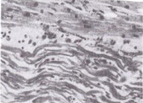
Coagulation necrosis (Figure 1) is the common pattern of necrosis characterized by cytoplasmic coagulation. Myocardial cells become acidophilic, but the features of myocardial fibers can be recognized for a long time from the onset of the pathologic process. However, the specific details of the structure change their properties. This type of necrosis commonly has to be recognized as a result of a vascular mechanism related to a reduced perfusion of specific areas of the myocardium, although other chemical, physical and biological factors may play their effects.

Figure 1 Coagulative necrosis of myocardium. The myocardial tissue under necrosis shows altered structure of muscular fibers, although with still evident composition. Leukocyte infiltration may be seen in the infarct area. Hematoxylin-Eosinstaining.
With regard to this type of necrosis, Katz14 & Schwartz15 hypothesized a loss of contractile properties of myocardial fibers as an effect of intracellular acidosis derived by the anaerobic glycolysis due to the hypoxia. Hypoxia should be responsible of a shift of Calcium ions and contractile myocardial protein coupling. There is evidence that cigarette smoking can biochemically determine these changes.16 These coagulate cells may meet a liquefactive process or be removed by fragmentation and phagocytosis due to macrophages and leukocytes migrating in the altered area (Figure 1).
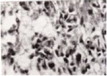
Colliquative myocytolysis (also named liquefaction necrosis) displays an evident liquefaction of the necrotic tissue due to the action of hydrolytic enzymes released from myocardial fibers undergone autolysis. They result from the migration of leukocytes in the ischemic area (Figure 2). Marked nuclear alterations with fragmentation, basophilia and degeneration characterize this type of necrosis, which determines myocardial lysis, vacuolization with loss of contractile proteins of the myocardium, and edema, all lesions that precede the development of heart failure.17 (Figure 2).

Figure 2 Colliquative myocytolysis. Evident vacuolization of the myocardium undergone lysis as a result of leukocyte infiltration. Hematoxylin-Eosin staining.
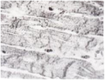
Coagulative myocytolysis (Figure 3) is undoubtedly the type of myocardial necrosis more closely related to the action of both smoking toxics, nicotine and carbon monoxide.18–20 The histologic features and pathogenic mechanisms can be easily recognized because being similar to that due to catecholamine release and sympathetic nervous system stimulation.21,22

Figure 3 Coagulative myocytolysis. Myocardial fibers are deeply altered and contractbands due to cell death in hypercontraction, similarly to what observed in the “stone heart”, may be seen. Hematoxylin-Eosin staining.
The main alterations that characterize coagulative necrosis consist of a variety of changes of myocardial fibers, which meet hypercontraction with the formation of transverse bands, fragmentation, and rupture. This picture evokes the pattern defined as “alveolar pattern”23 responsible of a strong myocardial disarray considered as an architectural disorganization of the myocardium. It has beenclosely linked with the adrenergic stress.24 It is worth noting that smoke cardiomyopathy takes a significant place in the context of the various types of myocardial necrosis.
This pattern is a result of a direct effect of carbon monoxide on myocardial cells and intracellular structures of the heart muscle.25–27 The lesions observed are an association of some features of the necrosis described and consist of hyalinosis, interstitial edema, perivascular cellular infiltrates and hemorrhagic foci resulting from myocardial fiber fragmentation. As can be seen a wide spectrum of alterations characterize myocardial necrosis making possible multiple patterns typical of the underlying disease. However, it can be also determined by cigarette smoking as experimentally documented. Therefore, even if it is hard to clearly attribute the different morphology of myocardial necrosis to tobacco toxics, nevertheless some of the alterations can lead to this way.
It is worth noting that a large number of pathogenic mechanisms can be attributed to the cigarette smoking, but, with regard to myocardial necrosis, only two are worthy of mention (Table 2): a toxic, and a coronarogenic mechanism.
|
Toxic mechanism |
Vascular mechanism |
|
Carbon monoxide (carboxyhemoglobin) |
Coronary atherosclerosis |
|
Nicotine (adrenergic and sympathetic stimulation) |
Coronary narrowing |
Table 2 Pathogenic mechanisms of myocardial necrosis from cigarette smoking
There is evidence that all infarcts of the heart muscle belong to the group of necrotic lesions, but not all cardiac necrosis are necessarily infarcts28 Starting from this assumption, the different types of necrosis described can be interpreted to have the same, but also different pathogenic mechanisms.
Vascular mechanism of necrosis primarily recognizes pathological alterations of the coronary arteries, which undergo narrowing and/or occlusion as a consequence of a thrombus formation. Morpho-pathological substrate is coagulation necrosis, which occurs because of reduced coronary blood flow and hypoxia due to the action of nicotine and carbon monoxide. The two pathogenic mechanisms are often associated. Thus, it has been well documented the effects of both nicotine, mainly by endothelial dysfunction,29,30 and carbon monoxide on the coronary tree. Increased concentrations of carboxyhemoglobin31 contributeto activating the vascular mechanism.
Toxic mechanism, mainly exerted by nicotine in the first phases of damage through an increased catecholamine release and sympathetic nervous system stimulation,32,33 and later as a direct effect of the hypoxia chronically caused by carbon monoxide,34,35 may be responsible of the different types of necrosis described, and primarily smoke cardiomyopathy.
There is no doubt that some chemical compounds of cigarette smoke, primarily nicotine and carbon monoxide, play a severely adverse effect on heart muscle cells causing a wide spectrum of structural lesions mainly of ischemic type. These involve both myocardial fibers and intracellular structure, and are able to determine different types of myocardial necrosis, which have been certainly documented in the experimental pathology of the animals.
None.
The authors declare no conflicts of interest.
None.

©2016 Leone. This is an open access article distributed under the terms of the, which permits unrestricted use, distribution, and build upon your work non-commercially.