Journal of
eISSN: 2373-4396


Case Report Volume 9 Issue 6
Loma Linda University Medical Center, USA
Correspondence: Howard Lan, Loma Linda University Medical Center, Loma Linda, CA, USA
Received: September 26, 2017 | Published: October 6, 2017
Citation: Lan H, Mamdani N (2017) The Use of Handheld Ultrasound in Early Diagnosis and Treatment of Life Threatening Cardiac Condition: A Case Report of Early Diagnosis and Treatment of Cardiac Tamponade. J Cardiol Curr Res 9(6): 00342. DOI: 10.15406/jccr.2017.09.00342
Ultraportable handheld ultrasound machines have been available in the health care market since 2010. There are wide array of uses for handheld ultrasound including applications in the field of obstetrics, emergency medicine, and cardiovascular imaging. Handheld ultrasound helps clinicians perform point-of-care bedside assessment and to make timely decisions which impact the outcome of patients. We present a case report demonstrating the use of handheld ultrasound helped with early diagnosis and treatment of cardiac tamponade.
A handheld ultrasound (HHU) is defined as a “pocket-size, hand-held imaging devices.”.1 The European Association of Echocardiography.1 gave eight recommendations for its use: 1. Complement to a physical examination in the coronary and intensive care unit, 2. Tool for a fast initial screening in an emergency setting, 3. Cardiologic counselling in or outside health-care facilities and hospitals, 4. First cardiac evaluation in ambulances, 5. Screening programs in schools, industry, and community activities, 6. Triaging candidates for a complete echocardiographic examination, 7. Teaching tool, 8. Semi-quantification of extravascular lung water. In literature, there are myriad uses of HHU that have been described including epidural analgesia for women in labor,2 evaluation of breast mass,3,4 evaluation of blood supply prior to plastic surgery,5 measurement of optic nerve sheath diameter to evaluate for intracranial pressure,6 detection of fetal growth restriction,7 evaluation of trauma patients,8 diagnosis of bone fracture,9 teaching cardiovascular anatomy to medical students,10 and evaluation for heart failure.11
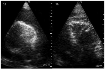
A 34 year-old male with history of hypertension, diabetes, and end stage renal disease on hemodialysis who was sent to the emergency room due to hypotension during outpatient hemodialysis. In the emergency room, patient was hemodynamically unstable. Patient was intubated for acute respiratory failure and was started on pressors due to shock. Patient’s cardiac exam showed diminished heart sounds and bedside echo performed by a HHU (GE Vscan®) showed a large pericardial effusion (Figure 1a). Other physical exam findings included pulsus paradoxus which confirmed cardiac tamponade. An emergent pericardiocentesis was performed at bedside, with the guidance of HHU, and 2.5L of pericardial effusion was removed. Repeat bedside echo showed trace residual pericardial effusion with expanded right ventricle filling (Figure 1b), and patient’s hemodynamics improved immediately.

Figure 1 a) Apical 4 chamber view in mayo format (RV on right side) showing large pericardial effusion and RV diastolic collapse.
b) Post 2.5L pericardiocentesis showing residual small size pericardial effusion with improved RV filling. Images were downlosded from GE VscanTM (Fairfield CT) handheld ultrasound.
With advancing technology in ultrasound, devices are now smaller, more portable, cheaper, and have improved interface with electronic medical record.12 Utilization of HHU for daily use in clinical practice might become universal over time. There is much debate on whether HHU will replace the stethoscope.13,14 however, most would agree that HHU augments physical findings at bedside.15 and could assist physicians in making important clinical decisions.
Transthoracic echocardiogram performed by HHU has been shown to be cost effective and could provide accurate detection of cardiac structural and functional abnormalities.16 When used by cardiologists, HHU provides a more accurate diagnosis than physical examination alone for the majority of common cardiovascular abnormalities.17 In this case report, the use of HHU to supplement the cardiac exam resulted in early diagnosis and treatment of cardiac tamponade, a medical emergency. In literature, there are two prior reports of cardiac tamponade that were diagnosed and treated with HHU guidance.18,19, both with good clinical outcome similar to our case.
There are variety of uses for HHU in the field of medicine. HHU is currently used as an adjunct to the standard physical examination at bedside. In this case report, the use of HHU supplementing the cardiac exam lead to early diagnosis and treatment of cardiac tamponade which resulted in immediate improvement in patient’s hemodynamics and outcome.
None.
None.

©2017 Lan, et al. This is an open access article distributed under the terms of the, which permits unrestricted use, distribution, and build upon your work non-commercially.