Journal of
eISSN: 2373-4396


Case Report Volume 4 Issue 1
Heart Center, Kaplan Medical Center, Affiliated to the Hebrew University, Israel
Correspondence: Jacob George MD, Heart Center, Kaplan Medical Center, Affiliated to the Hebrew University, P.O.B 1, Pasternak, Rehovot, Israel, Tel 972-8-9441335
Received: November 13, 2015 | Published: November 25, 2015
Citation: George J, Zahangir NM, Ahmed ST, et al. Sinus of valsalva fistula. J Cardiol Curr Res. 2015;4(1):119-122. DOI: 10.15406/jccr.2015.04.00132
A 56-year-old male presented with new onset of congestive heart failure symptoms and a continuous murmur. Transthoracic and transesophageal echocardiography showed severely reduced left ventricle function and shunt flow from an aneurysm in the non-coronary cusp of the aortic valve to the right atrium. The patient underwent surgery with 3 coronary artery bypass grafts and direct closure of the fistula with no residual flow.
Keywords: aortic valve aneurysm, aortic valve fistula, echocardiolgraphy, heart failure
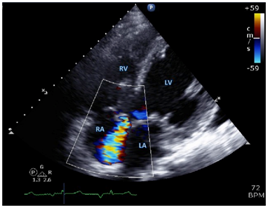
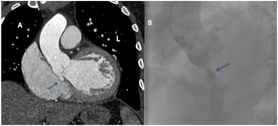
A 56-year-old man was admitted to the emergency department with effort dyspnea, fatigue and leg edema. The patient had a history of hypertension, diabetes mellitus and left ventricle hypertrophy and no family history of cardiac disease. The patient was fully alert with mild respiratory distress. The physical examination revealed sunken sternum, consistent with pectus ex cavatuma, crackles in the lower parts of the lung, pitting edema and a 3/6 continuous murmur in the left sternal border. The laboratory findings were unremarkable. The patient was treated in the emergency department with sub lingual nitrates and loop diuretics with marked improvement. Transthoracic echocardiography showed severely reduced left ventricle function with estimated ejection fraction of 20% and an echo-lucent tunnel of 6mm with flow from the aortic root to the right atrium (Figure 1). Trans-esophageal echocardiography confirmed the presence of left to right shunting from an aneurysm of the non-coronary cusp of the aortic valve to the right atrium (Figure 2). Cardiac CT and cardiac catheterization provided additional views of the fistula and demonstrated three-vessel coronary artery disease (Figure 3). The patient underwent cardiac surgery. During surgery, an opening measuring 7mm, partially covered with a membrane, was seen on the non- coronary annulus (Figure 4). The patient underwent a direct closure of the fistula with no residual flow in the fistula as observed by intra operative trans-esophageal echocardiography. Three coronary artery bypass grafts were also placed.

Figure 1 Transthoracic Echocardiographic Study. In the apical four-chamber view obtained at mid – systole with color Doppler Jet flowing from the aortic valve to the right atrium.

Figure 2 Trans Esophageal Echocardiographic Study. In the short axis view at the level of the aortic valve obtained at early–diastole with color Doppler we could locate the jet flow between the non-coronary cusp and the right atrium. (Panel A). Small magnification of the aortic valve without Doppler color reveals an aneurysm at the sinus of valsalva of the non-coronary cusp, approximately 10x10 mm in size, marked by asterisk.

Figure 3 Cardiac CT (Panel A) and Cardiac catheterization (Panel B).
The arrow in panel A points the location of the sinus of valsalva aneurysm, approximately one cubic centimeter in size. Please note that the color of contrast material is similar in the aorta and the aneurysm, signifying its origin. The arrow in Panel B points to the same structure. The contrast material fills the space and then fades into the chamber.

Figure 4 The aorta is cut while the patient is on cardiac bypass machine. The arrow points to the location of the aneurysm and resulting fistula, at the base of the non-coronary cusp of the aortic valve.
RA denotes right atrium, RV right ventricle, LA left atrium and LV left ventricle. RA denotes right atrium, RV right ventricle, LA left atrium, RCC right coronary cusp, LCC left coronary cusp, N non-coronary cusp and asterisk aneurysm.
Recognition of new physical finding, echocardiography and the use of different imaging modalities, allowed a fast and accurate diagnosis and treatment of new onset of heart failure.

©2015 George, et al. This is an open access article distributed under the terms of the, which permits unrestricted use, distribution, and build upon your work non-commercially.