Journal of
eISSN: 2373-4396


Case Report Volume 3 Issue 5
Chief of Cardiology, USA
Correspondence: Hugo Chinchilla, Chief of Cardiology, Military Hospital, Tegucigalpa, Honduras, El Ocotal, Francisco Morazan, USA
Received: December 13, 2014 | Published: October 12, 2015
Citation: Calix HC (2015) Revascularization in Multiple Vessels. J Cardiol Curr Res 3(5): 00114. DOI: 10.15406/jccr.2015.03.00114
It is male patient aged 60 with a history of Chronic hypertension, dyslipidemia, and Chronic smoker. 5years ago the patient had an aorto coronary bypass to present box angina class III of Canadian society of cardiology. 6months after surgery for coronary Revascularization presented symptoms of progressive angina to be why undergoes a percutaneous coronary intervention (PCI) of native vessels in 2times again class III (SCC), achieving complete Revascularization after placement of medicated sten (endeavor, Medtronic) zotarolimus-eluting. With excellent angiographic and Clinical outcome. The patient remains asymptomatic and makes normal life.
Keywords: coronary artery bypass grafting; percutaneous coronary intervention; class iii (nv) native vessels; drug-eluting sten
The patient is a 60year old man with a known history of Chronic hypertension controlled with enalapril 20mg per day and mixed uncontrolled dyslipemia. He doesnot have previous history of smoking or diabetes. Five years ago he under went cardiac bypass surgery that was complicated immediately with mediastin it is secondary to a wound infection over the sternum; for this reason he was admitted to the intensive care unit where he remained for two months. Six months after being discharged, he presented with anginal Chest pain with dyspnea that rapidlyprogressed to minimaleffortseven with intensive medical support. Wemethimfor the first time onNovember of 2007 when he had a positive stress test early (positive at 1minute 10seconds) in stage I of the BRUCE protocol with ST depressionanteriorly and inferiorly with a delayedrecoveryclinically and electrocardiographically.

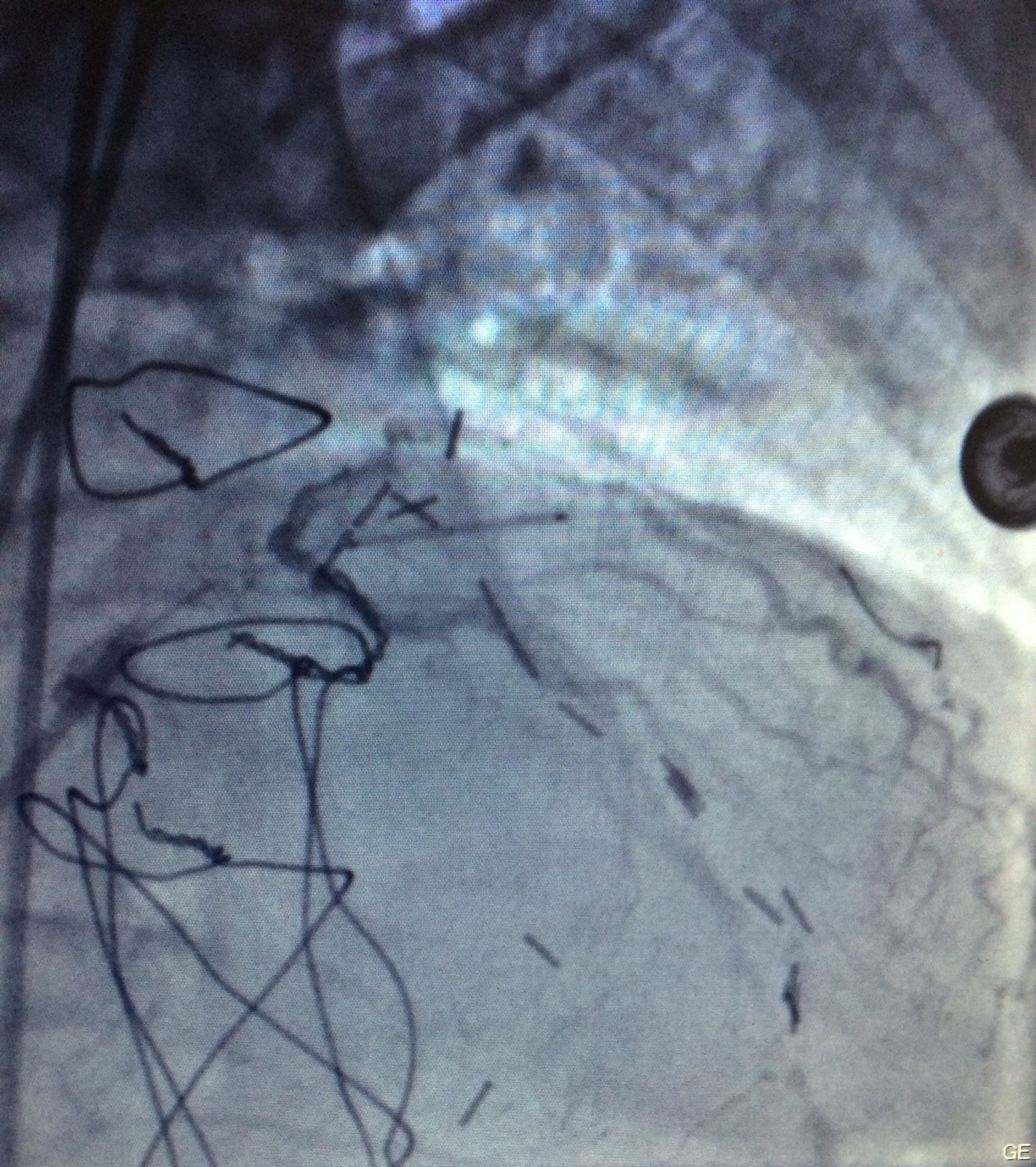
Figure 3 Through a conventional right femoral approach, the left coronary ostium was canalized using a 4.0 6Fr Launcher guiding catheter (Medtronic). Subsequently, a floppy microguide at the level of the anterior left descending artery was used to cross the lesion. Balloon dilation was performed to open the vessel starting with a 1.5x18mm Sprinter balloon (Medtronic) followed by a 2.0x22mm to finally deploy a 2.5x18mm Endeavor stent at 18atm for 10 seconds at the level of left anterior descending artery obtaining a satisfactory angiographic result.
Through a conventional right femoral approach, the left coronary ostium was canalized using a 4.0 6Fr Launcher guiding catheter (Medtronic). Subsequently, a floppy micro guide at the level of the anterior left descending artery was used to cross the lesion. Balloon dilation was performed to open the vessel starting with a 1.5x18mm Sprinter balloon (Medtronic) followed by a 2.0x22mm to finally deploy a 2.5x18mm Endeavorstent at 18 atm for 10seconds at the level of left anterior descending artery obtaining a satisfactory angiographic result (Figure 3-6).

Figure 4 Through a conventional right femoral approach, the left coronary ostium was canalized using a 4.0 6Fr Launcher guiding catheter (Medtronic). Subsequently, a floppy microguide at the level of the anterior left descending artery was used to cross the lesion. Balloon dilation was performed to open the vessel starting with a 1.5x18mm Sprinter balloon (Medtronic) followed by a 2.0x22mm to finally deploy a 2.5x18mm Endeavor stent at 18atm for 10 seconds at the level of left anterior descending artery obtaining a satisfactory angiographic result.

Figure 5 Five months later we decided to treat the ramus intermedius. We predilated the lesion with a 2.5x22mm Sprinter Balloon (Medtronic) to place a 2.5x22mm Endeavor stent inflated at 16 atm for 10 seconds obtaining good angiographic result.

Figure 6 Five months later we decided to treat the ramus intermedius. We predilated the lesion with a 2.5x22mm Sprinter Balloon (Medtronic) to place a 2.5x22mm Endeavor stent inflated at 16 atm for 10 seconds obtaining good angiographic result.
Five months later we decided to treat the ramus intermedius. We predilated the lesion with a 2.5x22mm Sprinter Balloon (Medtronic) to place a 2.5x22mm Endeavor stent inflated at 16 atm for 10seconds obtaining good angiographic result.
The patient is currently asymptomatic living a normal life. Stress tests were both clinically and electrically negative for myocardial ischemia with good physical capacity.
None.
Author declares there are no conflicts of interest.
None.

©2015 Calix. This is an open access article distributed under the terms of the, which permits unrestricted use, distribution, and build upon your work non-commercially.