Journal of
eISSN: 2373-4396


Case Report Volume 9 Issue 6
1Division of Cardiovascular Medicine, Clinic Hospital, Asuncion National University, Paraguay
2Department of Cardiovascular Surgery, Clinic Hospital, Asuncion National University, Paraguay
3Pahology Department, Health and Science Investigation Institute, Clinic Hospital, Paraguay
Correspondence: Osmar Antonio Centurión, Professor of Medicine, Asuncion National University, Chief, Division of Cardiovascular Medicine, Clínic Hospital, Dirección: Av. Mariscal López e/ Coronel Cazal. San Lorenzo, Paraguay
Received: October 08, 2017 | Published: October 25, 2017
Citation: García-Bello LB, Bedoya DM, Figueredo SJ, Centurión OA (2017) Refractory Heart Failure despite Optimized Medical Treatment Which Subsides only after Surgical Resection of a Large Left Atrial Myxoma. J Cardiol Curr Res 9(6): 00344. DOI: 10.15406/jccr.2017.09.00344
A 59-year-old woman with diabetes mellitus who presented with complaints of progressive dyspnea, orthopnoea, lung congestion, hepatomegaly, and inferior limbs oedema that began about 6 months earlier is presented. At the time of hospitalization, she was in New York Heart Association (NYHA) functional class IV in sinus rhythm. Despite optimization of the medical treatment installed for the signs and symptoms of her heart failure there was no improvement in her clinical condition. We are reporting a patient with refractory heart failure despite optimized medical treatment which completely subsides only after surgical resection of large left atrial myxoma.
Keywords: left atrial myxoma, refractory heart failure, surgical resection
Atrial myxoma (AM) is a primary cardiac tumor that mainly localizes in the left atrium. Primary cardiac tumors are infrequent (5%) and about 75% of them are benign. Half of these rare cardiac tumors are AM.1 The global annual incidence of AM is 0.5 per million people and 75% of them are located in the left atrium.2 The clinical presentation varies among patients and certainly depends on the size, mobility and location of the AM. It can be asymptomatic, although the typical symptoms involve systemic embolism, heart failure, or non-specific symptoms. Systemic embolism occurs in 25% to 50% of left AM and about half of them travel to the central nervous system.2,3 The clinical diagnosis of AM is based on imaging methodology like echocardiography, cardiac computerized tomography or cardiac magnetic resonance imaging. These imaging techniques provide detailed information about de structure of the tumor mass, its mobility, size, and myocardial invasion. The histopathological study of the cardiac mass confirms the diagnosis of AM.4 In order to avoid embolism and other cardiac and systemic complications total surgical resection of the intra-atrial myxoma should be done as soon as possible. Therefore, we are presenting this relatively uncommon disease of a left atrial myxoma developing refractory heart failure which subsides only after surgical resection of this intra-atrial mass.
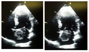
A 59-year-old woman with diabetes mellitus presented with complaints of progressive dyspnea, orthopnoea, inferior limbs oedema that began about 6 months earlier with milder symptoms. She denied previous consultation and treatment. At the time of hospitalization, she was in New York Heart Association (NYHA) functional class IV in sinus rhythm; her blood pressure was 140/90 mmHg. She had mild bilateral basal lung congestion and ascites, mild hepatomegaly and inferior limbs oedema was present. The first heart sound was increased. A soft mid-diastolic mitral murmur was present, as well as, a grade III/VI pan-systolic mitral murmur heard best at the apex and radiated to the axilla. The conventional electrocardiogram showed sinus rhythm. Transthoracic color-flow Doppler echocardiography revealed a hyper-echogenic large left intra-atrial mass (49 x 44 mm) suggesting an atrial myxoma which was attached to the inter-atrial septum (Figure 1). She also had moderate mitral regurgitation due to valve coaptation failure, mild to moderate left atrial dilatation (48 mm), and moderate pulmonary hypertension with a peak systolic pulmonary artery pressure of 47 mmHg. Mitral valve diastolic velocity was increased to 2.3 m/s with a mean pressure gradient of 10 mmHg. The LV ejection fraction was 69%. Blood tests were within normal limits. The carotid Doppler ultrasound, as well as, the coronary angiogram revealed arteries within normal limits. Despite optimized medical treatment installed for the signs and symptoms of her heart failure there was no improvement in her clinical condition.

Figure 1 Transthoracic color-flow Doppler echocardiography at four chamber apical view showing a hyper-echogenic large left intra-atrial mass suggesting an atrial myxoma which was attached to the inter-atrial septum. 1A: Shows the atrial myxoma almost filling the entire atrium during systole with the mitral valve closed. 1B: Shows the atrial myxoma during diastole with the mitral valve wide open.
After obtaining informed consent, the patient underwent successful surgical resection of the intra-atrial mass which consisted of a soft white and yellowish friable myxomatous mass that measured about 5 x 5.5 approximately (Figure 2) which was confirmed to be an atrial myxoma with histological studies revealing mixed stromal tissue (Figure 3). She had a favourable post-surgical evolution. The echocardiography before discharge showed complete resolution of the pulmonary hypertension and of the mitral regurgitation and left atrial dilatation.
We are reporting a case of a patient with refractory heart failure despite optimized medical treatment which completely subsides only after surgical resection of a large left atrial myxoma. Despite optimized medical treatment installed for the signs and symptoms of her heart failure there was no improvement in her clinical condition. Evidently the large and mobile left atrial myxoma was generating a mitral valve stenosis-like mechanical obstruction since she developed a moderate left atrial dilatation and moderate pulmonary hypertension which disappeared after resection of the large atrial mass.
The prevalence of intracardiac tumors is only 0,002%-0,3%. AM usually appear at the age of 30 to 60 years old.1-4 Atrial myxomas tend to differ in shape, size, and texture, and originate from the vicinity of the fossa ovalis in the inter-atrial septum in 75% of cases like in the patient that we are presenting here. The clinical and differential diagnosis of AM is based on imaging methodology like echocardiography, cardiac computerized tomography or cardiac magnetic resonance imaging.5-8 These imaging techniques provide detailed information about de structure of the tumor mass, its mobility, size, and myocardial invasion. The histopathological study of the cardiac mass confirms the diagnosis of AM. The surgical treatment consists of the total resection of the intra-atrial mass which should be prompt enough and complete to avoid embolism and other cardiac complications.4-6 When an AM is diagnosed with any imaging methodology it usually implies immediate consequent surgical excision to prevent embolic events. Therefore, studies with documented growth rate are very rare to find, and the actual growth rate remains a controversial issue. However, the calculated growth rate was estimated to be an average growth rate of 0.49 cm/month. Hence, the growth rate of atrial myxomas may be faster than is usually believed. The knowledge of this relatively fast growth of AM has clearly therapeutic implications. Surgery should be done as prompt as the diagnosis is made. Survival after total resection of the AM is high. Nevertheless, since there is a 1% to 3% recurrence long-term follow-up with echocardiography is highly recommended.1-3 With the advent of current auxiliary diagnostic methods, it is now feasible to do genetic studies and screen the relatives of patients having atrial myxomas to rule out additional occult familial cases and perform a surgical resection as early as possible to avoid cardiac and systemic complications in asymptomatic patients.
None.
There were no financial interest or conflict of interest.

©2017 García-Bello, et al. This is an open access article distributed under the terms of the, which permits unrestricted use, distribution, and build upon your work non-commercially.