Journal of
eISSN: 2373-4396


Case Report Volume 1 Issue 7
Consultant Pediatric Cardiologist, Al Qassimi Hospital, United Arab Emirates
Correspondence: Mahmoud Alsoufi, Consultant Pediatric Cardiologist, Al Qassimi Hospital Sharjah, United Arab Emirates, Tel 00971-553405476
Received: December 12, 2014 | Published: December 23, 2014
Citation: Alsoufi M. Heart block after ASD device closure.J Cardiol Curr Res. 2014;1(7):196-198. DOI: 10.15406/jccr.2014.01.00040
Transcatheter Device closure for secundum ASD is the treatment of choice when the patient has a good rims.1,2 and it has an excellent shore and long term results with minimal complications.3,4 Rhythm disturbance is one of these uncommon complications.5–7 We reported a 4 years old boy that developed a first and second degree heart block few days after device closure of large ASD secondum with spontaneous recovery after one week treatment with prednisolone.
Keywords: heart block, atrial septal defect asd, device closure, recovery, patient
4 years old boy with weight of 10.5Kg and height of 100cm, he is known case of moderate to large ASD secondum with recurrent chest infections and failure to thrive under treatment with frosimed and close follow up in the clinic. Cardiovascular examination reviled: normal S1+fixed and splitting of S2+ejection systolic murmur at left upper sternal border, per-procedure electrocardiogram showed normal sinus rhythm with heart rate 95-100/min ,incomplete right bundle brunch block. His echocardiography reviled large ASD secondum of 15-16 mm with adequate SVC, IVC and aortic rims and slightly short AV valve rim associated with dilated right atrium and ventricle with sings of increased pulmonary blood flow. During cardiac catheterization under general anesthesia and transesophaygeal echocardiography guidance we confirmed the accurate size of the defect and decided to precede and close this ASD by 18mm Amplatzer septal occluder (the device/height ratio is : 0.18).
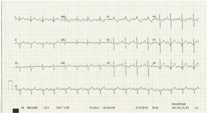
The procedure went smoothly without any complications in the cath lab and the patient was observed in the recovery area then in PICU with stable vital signs, sinus rhythm around 99-101/min and after 24h of the closure electrocardiography showed normal sinus rhythm with same degree of PRBBB (Figure 1). Echocardiography showed big ASD device insite without any residual shunt or interacting with adjacent structures (Figure 2). Patient was discharged home on aspirin 50mg daily and given appointment after one week for follow up in the clinic. After one week he came to pediatric cardiology for follow up, he was asymptomatic with no complain, his heart rate was 66/min with regular rhythm, other vital signs were stable. Echocardiography at that time showed ASD device in good position without any residual shunt or interfering with A-V valves or other structures. His ECG showed dominant sinus rhythm with HR=66/min , prolonged PR interval l0.18-0.2 sec (first degree heart block) and occasional 2:1 heart block (Figure 3).

Figure 1 ECG after 24 hour from the procedure showing normal sinus rhythm with partial RBBB same as before the procedure.
24hour holter monitor also confirm the finding of first degree heart block with occasional second degree as 2:1 but no escape rhythm or long pauses. The parents refused the admission for observation and they preferred to come daily for close follow up so we started him on oral prednisolone 1mg/kg/day with ECG monitoring every 2days. After 5days from this plan the patient remain stable with no complain but his ECG showed normal sinus rhythm with shorter PR interval 0.16-0.17 sec and no signs of heart block (recovered normal sinus rhythm) (Figure 4).
24hour holter monitor also confirm the normal sinus rhythm without any arrhythmias so we stopped the prednisolone and kept him under observation every week with new ECG. After one month he was stable, no complains and started to gain some weight on aspirin only.ECG at that time showed normal sinus rhythm with normal PR interval 0.16 sec and no signs of heart block (Figure 5). Echocardiography revealed the device in place, no residual shunt, no interfering with adjacent structures and no pericardial effusion.
ASD device closure usually is a procedure of choice for ASD secondom when the patient has good rims and files full the criteria for that.8–10 rhythm disturbance is very rare complication of this procedure,7 and usual common in small children who has large defects need large device for suitable closure. Patient who needs device size≥18mm and device/patient height ratio>0.18 are Carrie the higher risk factors. The pathophysiology of this rhythm disturbance after Large ASD device closure in not well known tell now,6,7 butit might be related to the pressure of the large device on the nearby conductive tissue mainly AV node or mild edema in that site related to micro inflammatory reaction after device implantation.
For this reason we recommend a routine ECG before the device closure just to have a baseline to notice any abnormality due to the ASD itself then another ECG and 24h holter monitor a 24hafter the procedure to detect the early new changes and later one another ECG/24h holter monitor after 1-2week from the procedure especially in those small patient with large ASD after a big device implantation. The role the treatment by corticosteroids as a trial to enhance AV conduction is a good suggestion in spite of till now no supportive controlled studies.7 However, I used it in my patient with post procedural first and second degree AVB with good result within days after the treatment. Still we have these questions: when we have to take the device out surgically? How many days we can observe the patients and are we sure that the rhythm will be back to normal after that or our patient needs a pacemaker?? (Figure 6).
ASD device closure stills the treatment of choice for the selected patient without major complications.1,3 Rhythm disturbance is one of the uncommon complication might be occur after days or weeks from the implantation of large device in small children necessitating a close follow up for those patient with frequent ECGs and holter monitor and most likely has a good prognosis with spontaneous recovery to sinus rhythm in the majority of the cases. A course of corticosteroid might be helpful in those causes.
None.
Authors declare that there are no conflicts of interest.

©2014 Alsoufi. This is an open access article distributed under the terms of the, which permits unrestricted use, distribution, and build upon your work non-commercially.