Submit manuscript...
Journal of
eISSN: 2373-4396

Cardiology & Current Research

Review Article Volume 3 Issue 2

Dual LAD – contemporary review

Eyvaz Abbasov,1 Soltan Manafov,1 Fuad Abdullayev2

1Department of Radiology, Azerbaijan
2Department of Cardiac Surgery, Azerbaijan

Correspondence: Eyvaz Abbasov, M Topchubasov Surgical Research Center, Sharifzadeh 196 str., Baku, Azerbaijan, Tel 994503229299

Received: July 12, 2015 | Published: July 31, 2015

Citation: Abbasov E, Manafov S, Abdullayev F. Dual LAD – contemporary review. J Cardiol Curr Res. 2015;3(2):1-6. DOI: 10.15406/jccr.2015.03.00095

Download PDF

Abstract

Dual LAD had been reported to occur with an incidence of 1% by Morettin1 as well as Spindola-Franco et al.,2 There is a lot of case reports on dual left anterior descending (LAD) coronary artery. The first description of a type IV dual LAD distribution was made in 1939, by Waterston et al.,3 in the case of Sir James Mackenzie, who had this type of distribution. By now six types of this anomaly have been described in the literature. But there is no any review article describing this anomaly in general, so we decided to write one.

Keywords: dual LAD, coronary anomaly

Abbreviations

LAD, left anterior descending coronary artery

Introduction

Dual LAD had been reported to occur with an incidence of 1% by Morettin1 as well as Spindola-Franco et al.,2 There is a lot of case reports on dual left anterior descending (LAD) coronary artery. The first description of a type IV dual LAD distribution was made in 1939, by Waterston et al.,3 in the case of Sir James Mackenzie, who had this type of distribution. By now six types of this anomaly have been described in the literature. But there is no any review article describing this anomaly in general, so we decided to write one.

According to the literature, incidence of the coronary artery anomalies in general population is approximately 1%.4 They are often not associated with clinical signs and symptoms, but some of them can lead to sudden cardiac death. According to the Sudden Death Committee of the American Heart Association, coronary anomalies are responsible for 19% of death in athletes.5 However, these anomalies are usually discovered as incidental findings at the time of coronary angiography or autopsy.

In this anomaly LAD is «divided» into two parts – proximal (short) and distal (long). Each of them comes from different sites and it determines a type of this anomaly. Usually short LAD gives rise to main septal perforator and main (first) diagonal. Dual LAD is usually a benign anomaly. No cases of sudden cardiac death related to it have been described. Spindola-Franco et al.,2 have proposed an angiographic classification for dual LAD in 1983, consisting of four types of LAD anomaly.
  1. Type I: Running in the anterior interventricular sulcus, the short LAD is generally the source of all the major proximal septal perforators. The long LAD also runs in the anterior interventricular sulcus, descending on the left ventricular side of it, and then reentering the distal anterior interventricular sulcus in order to reach the apex.
  2. Type II: The short LAD is the same as in Type I. The long LAD descends over the right ventricular side before reentering the anterior interventricular sulcus.
  3. Type III: The short LAD is consistent with that described in Types I and II. The long LAD travels intramyocardially in the ventricular septum.
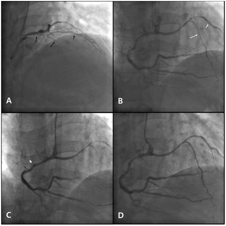
  4. Type IV: High in the anterior interventricular sulcus, a very short vessel is formed by the LAD proper and the short LAD. From this vessel, the major septal perforators, as well as the diagonal branches originate. The long LAD is unusual in its origin, arising from the right coronary artery (Figure 1 & 2). This classification was based on 23 cases. The long LAD arose from the LAD proper in 21 cases and from the right coronary artery in 2 cases. The initial course of the long LAD was on the epicardial surface of the left ventricle in 17 cases, right ventricle in 3 cases, and within the interventricular septum in 3 cases. A novel variant of dual LAD anomaly was described in 2010 by Machanda A et al.,6 They called it type V dual LAD. The short LAD was originating independently from the left coronary sinus and the long LAD was arising from the right sinus and had an intramyocardial course before reaching the distal interventricular groove. 

Figure 1 Coronary angiography and percutaneous coronary intervention.

  1. Short LAD (long black arrow) originating from the LMCA, giving rise to the D1 (short black arrow). There is significant stenosis in the proximal portion of the short LAD and the proximal portion of the D1.
  2. Long LAD (long white arrow) originating from the proximal RCA, coursing left, then turning downwards to the apex, giving rise to the D2 (long white arrow). There is significant stenosis of the distal RCA, the proximal PDA, the mid portion of the long LAD and the proximal portion of the D2.
  3. Deep engagement (arrowhead) with a 6 Fr MP guiding catheter to deliver stents to the distal RCA.
  4. Balloon angioplasty with a 6 Fr JR 4.0 guiding catheter at the mid portion of the long LAD and the proximal portion of the D2.

LAD: Left Anterior Descending Artery; LMCA: Left Main Coronary Artery; D1: First Diagonal Artery; RCA: Right Coronary Artery; PDA: Posterior Descending Artery; D2: Second Diagonal Artery; 6 Fr: 6 French; MP: Multipurpose; JR: Judkins Right.
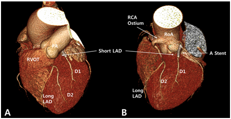
Figure 2 Computed tomographic coronary angiography.

  1. LAO cranial view showing the dual LAD anomaly with the short LAD on the proximal AIVG: giving rise to the D1 and the long LAD entering the mid AIVG giving rise to the D2.
  2. LAO cranial view with the RVOT removed: showing the short LAD originating from the left main coronary artery and the long LAD originating from the proximal RCA. The proximal RCA is taking off above the RCS and the long LAD is branching from the proximal RCA: passing between the RVOT and the aortic root. A stent can be observed from the proximal portion of the short LAD to the D1. The D2 is the dominant artery in the anterior wall of the left ventricle.

LAO: Left Anterior Oblique; LAD: Left Anterior Descending Artery; LMCA: Left Main Coronary Artery; RCA: Right Coronary Artery; AIVG: Anterior Inter Ventricular Goove; D1: First Diagonal Artery; D2: Second Diagonal Artery; RVOT: Right Ventricular Outflow Tract; PA: Pulmonary Artery; RoA: Root of Aorta.

In the case presented by Maroney J & Klein LW,7 they described another new variant (type VI) of dual LAD. The long LAD arose from the right coronary artery and followed a unique route underneath the right ventricular outflow tract in the interventricular septal area to the interventricular groove. Some authors think that this type may have greater clinical significance than other types because compression of the coronary artery between the right ventricular outflow tract and the aortic root in situations of increased pulmonary blood flow could cause coronary blood flow restriction and sudden cardiac death.8,9

Conclusion

As we may see, there are numerous variants of long LAD course. And it is possible that more types of this anomaly will be described in the future. But all of them have in common LAD territory supplied by two different vessels.

Acknowledgments

None.

Conflicts of interest

Author declares there are no conflicts of interest.

Funding

None.

References

  1. Morretin L. Coronary arteriography: uncommon observations. Radiol Clin North Am. 1976; 14(2):189˗208.
  2. Spindola-Franco H, Grose R, Solomon N. Dual left anterior descending coronary artery: Angiographic description of important variants and surgical implications. Am Heart J . 1983;105(3):445˗455.
  3. Waterston D, Orr J, Capell DF. Sir James Mackenzie's heart. Br Heart J. 1939;1(3):237˗248.
  4. Yamanaka O, Hobbs RE. Coronary artery anomalies in 126, 595 patients undergoing coronary arteriography. Cathet Cardiovasc Diagn. 1990;21(1):28˗40.
  5. Maron BJ, Thompson PD, Puffer JC, et al. Cardiovascular preparticipation screening of competitive athletes: A statement for health professionals from the Sudden Death Committee (Clinical Cardiology) and Congenital Cardiac Defects Committee (Cardiovascular Disease in the Young), American Heart Association. Circulation. 1996;94(4):850˗856.
  6. Manchanda A, Qureshi A, Brofferio A, et al. Novel variant of dual left anterior descending coronary artery. J Cardiovasc Comput Tomogr. 2010;4(2):139˗141.
  7. Maroney J, Klein LW. Report of a new anomaly of the left anterior descending artery: type VI dual LAD. Catheter Cardiovasc Interv. 2010;80(4):626˗629.
  8. Lee Y, Lim YH, Shin J, et al. A case report of type VI dual left anterior descending coronary artery anomaly presenting with no-ST-segment elevation myocardiac infarction. BMC Cardiovasc Disord. 2012;12:101.
  9. Angelini P. Coronary artery anomalies an entity in search of an identity. Circulation. 2007; 115(10):1296˗1305.
Creative Commons Attribution License

©2015 Abbasov, et al. This is an open access article distributed under the terms of the, which permits unrestricted use, distribution, and build upon your work non-commercially.