Journal of
eISSN: 2373-4396


Case Report Volume 1 Issue 2
1Department of Cardiology, Kosuyolu Kartal Heart Training and Research Hospital, Turkey
2Department of Cardiology, Kars Kafkas University, Turkey
Correspondence: Macit Kalcik, Department of Cardiology, Kosuyolu Kartal Heart Training and Research Hospital, Esentepe Mah, Milangaz Caddesi Unluer Sitesi B-Blok, No:22, Kartal/Istanbul, Turkey, Tel 90-536-4921789, Fax 90-216-4596321
Received: June 11, 2014 | Published: June 19, 2014
Citation: Kalcik M, Yesin M, Gursoy MO, et al. Delineation of a paravalvular leak origin between a rigid mitral ring and supra-annular implanted mechanical mitral prosthetic valve. J Cardiol Curr Res. 2014;1(2):53-54. DOI: 10.15406/jccr.2014.01.00011
Paravalvular leakage is not a rare condition among patients with mitral prostheses. Two-dimensional echocardiographic approaches are not sufficient to determine the origin of paravalvular leak that occurs after prosthetic mitral valve replacement. Real-time three-dimensional transesophageal echocardiography provides detailed structural identification of paravalvular leak origin and defect morphology compared to two-dimensional transesophageal echocardiography. Here we present a patient with paravalvular mitral regurgitation from a slit like defect between the mitral anular ring and the prosthetic valve delineated with the utility of 3D full volume and 3D color flow imaging modalities.
Keywords: mitral ring, paravalvular leak, transesophageal, echocardiography
TEE, Transesophageal echocardiography; 2D, two dimensional; 3D, 3 dimensional
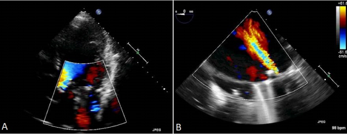
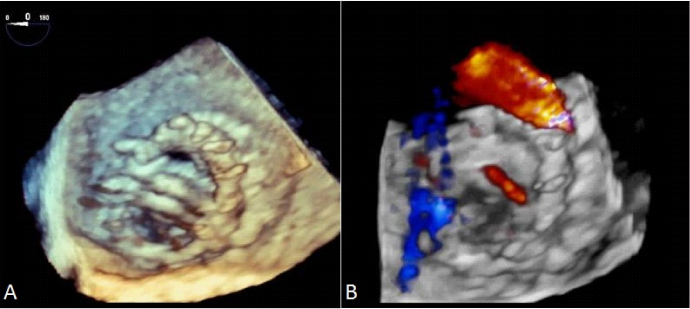
A 65year-old woman suffering from severe mitral valve regurgitation underwent mitral ring annuloplasty (31 no Saint Jude Medical Rigid Ring). After weaning from cardiopulmonary bypass pump, intraoperative transesophageal echocardiography (TEE) was performed this revealed withstanding severe mitral regurgitation. Therefore a supraannular 28 no bileaflet ATS-AP mechanical mitral valve was implanted over the mitral ring and intraoperative TEE showed normally functioning mitral prosthesis. The surgery was completed without complication. The patient experienced an uneventful postoperative period but was readmitted to hospital with ongoing NYHA Class 2 dyspnea one month after discharge. Transthoraic echocardiogram did not show any pericardial effusion but a moderate mitral regurgitation jet detected by color Doppler imaging (Figure 1A). TEE was performed to depict the origin of the regurgitant jet. Two dimensional (2D) TEE showed a moderate paravalvular leakage with the help of color Doppler imaging (Figure 1B). Subsequently, real-time 3 dimensional (3D) TEE revealed a regurgitates jet arising from a slit like defect between the mitral anular ring and the prosthetic valve with the utility of 3D full-volume and 3D color flow imaging modalities (Figures 2A & 2B).


Paravalvular leakage is not a rare condition among patients with mitral prostheses1 but a leakage between a mitral ring and mechanic prosthetic valve has not been reported so far. Real-time 3D TEE provides detailed structural identification of paravalvular leak origin and defect morphology compared to 2D TEE.2–5
None.
Authors declare that there are no conflicts of interest.

©2014 Kalcik, et al. This is an open access article distributed under the terms of the, which permits unrestricted use, distribution, and build upon your work non-commercially.