Journal of
eISSN: 2373-4396


Case Report Volume 5 Issue 6
King Saud bin Abdulaziz University for Health Sciences, King Abdulaziz Medical City, Saudi Arabia
Correspondence: Saad Al Bugami, King Saud bin Abdulaziz University for Health Sciences, King Abdulaziz Medical City, King Faisal Cardiac center, Jeddah 21423, Saudi Arabia
Received: May 13, 2016 | Published: May 24, 2016
Citation: Bugami SA, Althobaiti MW, Alrahimi J, Alsaiedi AJ, Kashkari WA (2016) Coronary Intervention of An Anomalous Left Main Coronary Artery Arising from the Right Sinus of Valsalva Presented as Acute Coronary Syndrome. J Cardiol Curr Res 5(6): 00184. DOI: 10.15406/jccr.2016.05.00184
Anomalous origin of the left coronary artery (LCA) from the right sinus of Valsalva is a rare congenital Anomaly. We report a 38 year old male presented with acute coronary syndrome (ACS). Referred to the Catheterization laboratory for Percutaneous Coronary Intervention (PCI). The coronary angiogram showed the LCA originating from the right coronary sinus. A critical stenotic lesion was observed in the shaft of the Left main coronary artery. CT coronary angiography displayed a benign retro aortic course. The lesion was treated successfully with stenting.
Keywords: percutaneous coronary intervention, ct angiography, anomalous coronary artery, congenital coronary artery
Congenital anomalies of the coronary artery are rare it ranges between 0.2-1.3percent.1–5 Anomalous origin of the coronary arteries is considered an incidental finding without clinical significance; however, these abnormalities may be responsible for angina pectoris, acute coronary syndrome, heart failure and increased risk of sudden death.6 It is important to define the course of these anomalies and exclude malignant ones especially if intervention is contemplated.7 We describe a young patient with anomalous left coronary artery arising from the right coronary cusp who presented with acute coronary syndrome and the way he was treated.
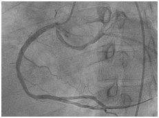
38 year old male patient with 15 packs per year smoking history presented to a peripheral hospital with acute onset typical chest pain for 30 minutes. His ECG revealed ST- Elevation acute lateral wall myocardial infarction. He was thrombolyzed with t-PA with complete resolution of his ECG changes. Echocardiogram showed mild LV dysfunction with LVEF of 45% and lateral wall hypo kinesis. He was then transferred to our center where he was taken to the catheterization laboratory and diagnostic coronary angiogram was done following obtaining an informed consent. It was difficult to engage the left coronary artery with Judking left catheter. A right judkins was taken to visualize the right coronary system. This revealed that the right coronary artery to be free of disease (Figure 1) and the left coronary artery to originate from the right coronary sinus. The anomalous left main showed a mid-shaft hazy and eccentric lesion (Figure 2), Normal left anterior descending and normal left circumflex. The patient was discussed in a heart team meeting and it was decided to obtain coronary CT angiogram to define the course of the anomalous left coronary artery which was retro-aortic (Figure 3). The patient was offered bypass surgery of which he declined to accept. The patient informed decision was to opt for PCI. A straight forward intervention was carried out using Judging’s 4 guiding catheter, which easily incubated the anomalous left main a BMW wire easily passed to the distal LAD and a direct 4.0x22 mm resolute stent deployed (Figure 4). A second stent 4.0x8 mm resolute stent deployed proximally to cover an area of plaque shift. A final IVUS was done which showed well stent apposition (Figure 5). The procedure was supplanted with 300mg of Plavix and 7000 unit of heparin. He was discharged next day.

Figure 1 Coronary angiography showing right and left coronary arteries originating from the right cornary sinus of Valsalva.
Anomalous origin of the left coronary artery from the right sinus of Valsalva is an uncommon malformation, accounting for 0.15% of cases in the series of Angelina et al.8 this anomaly is further subdivided in to 4 subtypes depending on the course it follows.1 The left main coronary artery courses between the aorta and the pulmonary artery.2 The left main coronary artery tracks anteriorly over the right ventricular outflow tract.3. The left main coronary artery takes an intra myocardial course before resurfacing at the proximal portion of the inter ventricular groove.4 The left main coronary artery passes posteriorly around the aortic root.8 Of these; the first anomalous configuration is classically considered the most dangerous placing patients at the highest risk of sudden cardiac death.9
It has been reported that anomalous coronary arteries are prone to atherosclerosis.10–12 The coronary blood flow would be disturbed in anomalous coronary arteries originating from the opposite side coronary sinus, which is located between the pulmonary trunk and the ascending aorta.13–16 Many patients with anomalous LCA arising from the right coronary sinus die before the age of 20 years and usually during or shortly after vigorous exertion.17–19 However, our patient did have a significant atherosclerotic changes in the anomalous Left main coronary artery. It appears that the cause of ACS in this patient might be related to coronary artery risk factors such as smoking rather than to the anomalous coronary artery itself which was found to have a more benign retro-aortic course.
We report a case with the retro aortic type of anomalous origin of the left coronary artery from the right sinus of Valsalva, this variant has been reported to have a relatively benign clinical course. The lesion in the left main coronary artery was thought to be due to atherosclerosis. It was decided to treat this patient percutaneously with direct stmplantation.PCI in anomalous coronary arteries is a feasible therapeutic strategy; however, accurate topographic identification of the origin and proximal course of the anomalous vessel is of paramount importance before proceeding in proceeding with these interventions.20
None.
The authors declare no conflicts of interest.
None.

©2016 Bugami, et al. This is an open access article distributed under the terms of the, which permits unrestricted use, distribution, and build upon your work non-commercially.