Journal of
eISSN: 2373-4396


Opinion Volume 17 Issue 3
1Department of radiodiagnosis and imaging, postgraduate institute of medical education and research, Chandigarh, India
2Department of cardiology, advanced cardiac centre, postgraduate institute of medical education and research, Chandigarh, India
Correspondence: Dr. Manphool Singhal, Professor, Department of Radiodiagnosis and Imaging, PGIMER, Chandigarh, India, Tel +91 9417308300
Received: June 21, 2024 | Published: June 28, 2024
Citation: Sharma A, Ghuman G, Paul S, et al. Common ostial origin of right subclavian artery and bicarotid trunk in two branch vessel left aortic arch-Rare aortic arch branching pattern. J Cardiol Curr Res. 2024;17(3):76-77. DOI: 10.15406/jccr.2024.17.00608
Varied anatomy has been described for the two branch vessel left sided aortic arch, most common being the brachiocephalico-carotid trunk in which left common carotid artery shares common origin with brachiocephalic trunk (also referred as bovine arch- actually a misnomer). We hereby describe a rare variant (bicarotico-right subclavian artery trunk) of two branch vessel left sided aortic arch in which there is presence of a bicarotid trunk which in turn shares common ostia with right subclavian artery (SCA) and separate origin of left SCA from the arch.
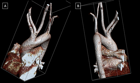
A 16 year old female came with complaints of change in voice for the past 2 months. There were also complaints of gradually progressing dysphagia to solids and not to liquids, dyspnoea on lying down. She underwent contrast CT scan of the chest which revealed a posterior mediastinal mass. Incidentally, a unique form of aortic branching pattern was seen i.e. two branch vessel pattern in a left sided aortic arch with common ostial origin of right SCA and bicarotid trunk (Figure 1).

Figure 1 Volume rendered CT images (A-B) showing common origin of right subclavian artery and bicarotid trunk in two branch vessel left aortic arch. 1-right subclavian artery. 2-bicarotid trunk, 3-right common carotid artery, 4-left common carotid artery, 5-left subclavian artery.
The development of aortic arch is a sequentially programmed complex process. Embryologically, six paired arches connect dorsal and ventral aorta. The aortic arch branches are formed by third, fourth and sixth arches. In normal branching pattern of left sided arch, the first branch is the right brachiocephalic artery followed by left common carotid artery and left SCA.
Any alterations in the above mentioned process will lead to various aortic arch anomalies, having prevalence of about 1-2% in the general population.1,2 Various classification systems were devised for aortic arch variants. In 2009, K. Natsis et al. classified aortic arch into eight types, based on the incidences recorded with type I being the most common and type VIII being the least common, using 633 digital subtraction angiographies in Caucasian greek patients.3 Recently in 2021, a systematic sub-classification of the left sided aortic arch variants was formulated based on cadaveric studies.4 Anomalous patterns were classified into types using the number of emerging branches (major as well as minor) and to the prevalence they appear. There are five subtypes of two branch vessel left sided aortic arch (as seen in the present case), most common out of which is the brachiocephalic-carotid trunk. However, the two vessel branching pattern in our case does not match with any of the described subtypes. In this, there is presence of a bicarotid trunk which shares a common origin with the right SCA and a separate origin of left SCA.
Bicarotid trunk represents the common single trunk of both the common carotid arteries. It is often associated with presence of aberrant right SCA.5 However, to the best of our knowledge, its common origin with right subclavian artery has not been described so far.
Meticulous knowledge of aortic variations plays a pivotal role in the management of patients by interventional radiologists, cardiologists, anaesthetists and surgeons. It allows accurate and early catheterization of arteries during angiographic studies. Moreover, it helps in choosing optimal cannulation and surgical strategy. Also, certain patterns like bovine arch has been linked with thoracic aortic dilatation and a risk of torsion and deceleration injuries considering only two fixation points in place of usual three points in normal branching pattern.6,7 Present case also highlights a rare two branch vessel left aortic arch along with highlighting the utility of CT in incidental detection of such unique variations.
None.
Authors declare no conflicts of interest.
Authors confirm that the manuscript is solely submitted to the journal and not submitted/ being considered anywhere else.

©2024 Sharma, et al. This is an open access article distributed under the terms of the, which permits unrestricted use, distribution, and build upon your work non-commercially.