Journal of
eISSN: 2373-4396


Clinical Paper Volume 16 Issue 5
1Department of Radiodiagnosis and Imaging, Postgraduate Institute of Medical Education and Research, Chandigarh, INDIA
22 Department of Cardiology, Postgraduate Institute of Medical Education and Research, Chandigarh, INDIA
Correspondence: Arun Sharma, MD, DM, Department of Radiodiagnosis and Imaging, Postgraduate Institute of Medical Education and Research, Sector-12, Chandigarh 160012, India, Tel 91 9910380907
Received: December 20, 2023 | Published: December 29, 2023
Citation: Manphool S, Sreenivasulu D, Arun S, et al. Bilateral persistent sciatic arteries: Rare vascular developmental anomaly. J Cardiol Curr Res. 2023;16(5):145-146. DOI: 10.15406/jccr.2023.16.00594
A 28-year-old female presented with non-specific complaint of persistent fatiguability. No history of claudication or signs of limb ischemia were present. On clinical examination, she was found to have absent bilateral femoral pulses, however the bilateral popliteal, posterior tibial and anterior tibial arteries were palpable. No obvious pulsatile swelling was appreciated in the bilateral lower limbs. Blood pressure in both lower limbs was within normal limits.
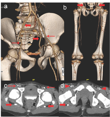
CT angiographic evaluation of the bilateral lower limbs demonstrated internal iliac arteries continuing as sciatic arteries (exiting though greater sciatic foramen with a posterior course) till popliteal fossa and dividing into normal anterior tibial artery and tibio-peroneal trunk. No associated aneurysm, stenosis or calcific plaques were noted. External iliac arteries were continuing as profunda femoris and incomplete superficial arteries (Figure 1). Findings were in keeping with complete bilateral persistent sciatic arteries with incomplete femoral arteries (Type III of Ahn-Min classification). The findings were discussed with the patient and no treatment was offered as there were no signs of limb ischemia or any associated abnormalities.

Figure 1 CT Angiography volume rendered images in anterior (a) and posterior projections (b) showing bilateral persistent sciatic arteries continuing as internal iliac arteries and coursing through the bilateral sciatic foramina (thick arrows) which are supplying the entire vascular axis of the leg (complete SPAs). The external iliac artery is continuing as CFA (thin arrows) which are continuing as incomplete superficial and profunda femoris arteries. Axial CT angiographic images at level of femoral head (c) and neck of femur (d) show posterior course of PSAs in the gluteal regions (thick arrows). The external iliac (thin arrows) and the profunda femoris (arrowheads) arteries are anterior and normal in course.
Persistent sciatic artery (PSA) is a rare congenital anomaly of the lower limb with reported incidence of 0.025%-0.04% and bilateral PSAs are still rarer accounting for only 12%-32% of all reported cases.1 Sciatic arteries supply to the lower limbs till the 6 weeks of gestation. As the fetus develops, sciatic artery involutes and persist as inferior and superior gluteal arteries, while profunda femoris artery supplies deep muscles of the thigh. PSAs results from the failure of regression of the sciatic artery and continues as internal iliac artery.1,2 Previously PSAs were classified into five types (Pillet classification) according to completeness of PSAs and superficial femoral arteries (SFAs) which was modified by Gauffre. Currently Ahn-Min's classification is in practice and classifies based upon on completeness of PSAs and SFAs along with associated symptoms and aneurysms.3
PSAs are usually asymptomatic in young and diagnosis is serendipitous. However, PSAs after 40 years of age can present clinically with pain when the artery lies within the sciatic nerve sheath, limb ischemia in 31-63%, thrombo-embolic complications leading to amputation in 8-10%, and with aneurysms occurring in up to 45% of cases.3-4
CT angiography optimally evaluates and helps in the accurate diagnosis of PSAs, evaluation of vascular supply to the limbs, complications and follow-up. Treatment strategies are customized according to presentation and abnormalities and range from medical management while aneurysms and their complications require either surgical or endovascular treatment.3-6
None.
No funding was received for the article..
Authors declare that there is no conflicts of interest/s to declare.

©2023 Manphool, et al. This is an open access article distributed under the terms of the, which permits unrestricted use, distribution, and build upon your work non-commercially.