Journal of
eISSN: 2373-4396


Clinical Paper Volume 16 Issue 5
Rivero-Covre Clinic Service, Villa Gdor Galvez, Medical Imaging Center, Rosario, Argentina
Correspondence: Mariana Pierucci Bosso, Rivero-Covre Clinic Service, Villa Gdor Galvez, Medical Imaging Center, Rosario, Argentina
Received: December 15, 2023 | Published: December 29, 2023
Citation: Bosso MP, Araceli MS. Apical microaneurysm as the only finding of chagasic heart disease. J Cardiol Curr Res. 2023;16(5):141-143. DOI: 10.15406/jccr.2023.16.00593
The characteristic macroscopic lesion and current pathological criterion of chronic Chagas cardiomyopathy is weight loss with segmental dyskinesia. Sometimes in the form of a "notch or niche", other times constituting a true "aneurysm". It has an incidence of 2-8.6% in asymptomatic patients and 24-64% of patients with myocardial involvement. The most frequent location is in the apex of the left ventricle 82%, 9% in the apex of the right ventricle and 9% in both.1 The pathophysiological mechanism is unknown; it could be caused by cardiac parasympathetic denervation generated by Trypanosoma cruzi during the acute phase of the disease.2 However, certain Chagas patients in indeterminate phase have aneurysms without presenting cardiac parasympathetic alterations.3-5 Other proposed theories are the greater mechanical stress that the myocardium must withstand, or that these segments are more vulnerable to certain pathogens.
The presence of a ventricular aneurysm is a predictor of thrombus formation, stroke and ventricular arrhythmia, so its diagnosis is essential.
Current guidelines recommend the ECG as the first study to be performed. The absence of ECG alterations would rule out cardiomyopathic involvement. Echocardiogram is of vital importance to detect cardiac damage, however aneurysms are a challenge since frequently, due to their location, they cannot be included within the ultrasound field. MRI is currently class IIb in the subclinical stage.6
Keywords: apical microaneurysm, chagasic cardiomyopathy
Clinical case: Patient aged 50 years, smoker (20 cig/day), sedentary, positive serology for recently diagnosed Chagas disease (6 months). He consulted for atypical precordial pain. Physical examination: BP 120/85 mmHg, HR 60 bpm, rest without particularities.
ECG: RS, 60 bpm, PR 160 msec, QRS 80 msec, QT 400 msec, Axis +30°, no ST-T segment alterations, no arrhythmias.
PEG: Stopped by exhaustion of MMII at 85% of the FCMT, No changes in ST-T, Frequent EV, monomorphic at maximum effort.
ECHO: Left and right cavities within normal parameters. Preserved LV systolic-diastolic function. At the level of the infero-apical region there is an evagination of the small ventricular cavity that seems not to present contractility, of difficult evaluation (it could correspond to a ventricular aneurysm by the patient's history), so it is suggested to complement with another diagnostic method (CMR with gadolinium), for its correct visualization and diagnosis. In the rest of the segments no parietal motility disorders were observed at rest.
RESO: Left ventricle with preserved volumes and mass. At the apex level there is a microaneurysm (saccular evagination, "gloved finger", of the myocardial wall, which is severely thinned: 1.5mm and dyskinetic). Normal parietal motility of the rest of the segments. Preserved systolic function. Late enhancement sequences do not show gadolinium uptake.
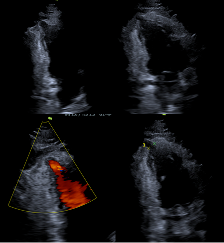
Imaging study: Figures 1-4.

Figure 1 Doppler echocardiogram, apical 2-chamber view: At the level of the apex there is evidence of evagination in the dyskinetic myocardial wall and apparent thinning, with evidence of color filling inside, neck measured 0.95 cm.

Figure 2 2-chamber echocardiography sequence: At the apex level, the presence of a microaneurysm (saccular evagination, "gloved finger", of the myocardial wall, which is severely thinned: 1.5mm and dyskinetic) is visualized.
Diagnosis of Chagas disease is based on epidemiology, positive serology, and clinical and imaging findings.
Disease stratification is determined by the degree of cardiac involvement and symptoms of heart failure.
Typical imaging findings include areas of segmental hypokinesia, aneurysms, fibrosis or mural thrombi, most commonly in the apex of the left ventricle.
ECG is the first study to be performed due to its low cost and availability. The absence of ECG alterations would rule out myocardiopathic involvement. However, 8.5% of asymptomatic patients present ventricular aneurysms, so it is reasonable to perform an echocardiogram in all patients with positive serology.
In our case the patient had positive serology for Chagas disease, was asymptomatic and with ECG without alterations (indeterminate phase), however imaging studies, echocardiography and MRI, allowed to recategorize it demonstrating the presence of apical aneurysm.
The diagnosis, risk stratification and management of patients with Chagas disease depends on myocardiopathic involvement, arrhythmogenic substrate and thromboembolic risk.
The accuracy of 2D and 3D echocardiography is very good for determining biventricular volumes and function; however, the presence of apical aneurysms is a challenge, since their location often prevents them from being included in the ultrasound field. The use of contrasts for LV opacification can be useful, although their availability is very limited.
Advanced echocardiographic techniques (global longitudinal strain, speckle traking) and the detection of early tissue changes have a clinical impact yet to be defined in the prediction of disease progression.7,8 Global longitudinal strain is the most validated method for the detection of subclinical LV functional damage in patients with Chagas disease and has a high correlation with the amount of myocardial fibrosis assessed by MRI.
Cardiac magnetic resonance imaging, thanks to its excellent spatial resolution and its capacity for tissue characterization, provides pathophysiological information on the disease. In 8% of patients with positive serology without ECG or ECHO alterations, fibrosis is present, which correlates inversely with LV systolic function and is a mechanism in the generation of ventricular arrhythmias. In addition, it is useful for assessing cardioembolic risk. Intracavitary thrombi imply a high risk of stroke and peripheral embolism, which may not be detected by echocardiography, even with the use of contrast agents.
None.
I declare there is no conflict of interest.

©2023 Bosso, et al. This is an open access article distributed under the terms of the, which permits unrestricted use, distribution, and build upon your work non-commercially.