Journal of
eISSN: 2473-0831


Research Article Volume 4 Issue 5
Correspondence: Susheel Kumar Mittal, Thapar University, Patiala, Punjab-147004, India, Tel 91 9815653261
Received: April 17, 2017 | Published: May 2, 2017
Citation: Mittal SK, Sharma R, Narang P (2017) A Green Electroanalytical Method for the Determination of Levofloxacin by Ion-Pair Formation with Picric Acid. J Anal Pharm Res 4(5): 00116. DOI: 10.15406/japlr.2017.04.00116
Levofloxacin (LFX), an antimicrobial agent has been detected by using picric acid (PA) as an anionic reagent in aqueous medium. On mixing the reactants in distilled water yellow colored product is separated within two minutes which was found to be an ion-association complex (LFX-PA) of both the reactants. Spectroscopic and voltammetric studies were carried out to explain the interaction between levofloxacin and picric acid. Spectroscopic study of the ion-pair product confirmed its formation as determined from substantial shift in respective λmax values. In voltammetric experiments, a shift of 70 mV was noticed in an anodic peak of LFX-PA (1.64V) accompanied with a large decrease in peak current. In addition, a number of new peaks appeared in voltammogram of LFX-PA. Similarly, in case of cathodic cycle, new peaks appeared in differential pulse voltammogram of the ion-pair which proved the interaction of the PA and LFX. A calibration curve was drawn using the peak current values of the LFX. The linear increase in peak current with increasing LFX concentration was obtained in the concentration range 1.5×10⁻³ M–6.0×10⁻³ M with p>=0.994. A study on the solvent effect on the spectrometric as well as voltammetric methods for LFX suggested that water is the best medium for the study. Stability of the complex was also determined by computational methods using DFT/B3LYP/6-31G basis sets. Further, analysis of LFX in commercially available drug tablets proved that the proposed method is valid in real life sample analysis.
Keywords drug analysis, dft calculations, ion-pair formation; picric acid, voltammetry
PA, picric acid; LFX, levofloxacin; FTIR, fourier transform infrared
Fluoroquinolones are a group of compounds which contain fluorine at 6-position of basic quinolone core structure. These are used for treatment of bacterial infection, pneumonia, urinary tract infection, joint and bone infection, skin infections, typhoid fever and several other infection conditions 1. Fluoroquinolones include Ciprofloxacin, Levofloxacin, Norfloxacin, Gatifloxacin and Ofloxacin. Other than core structure, these molecules also resemble in physicochemical properties, pharmacokinetic characteristics and microbial activities.
Structure of Levofloxacin
Levofloxacin, (−)-(S)-9-fluoro -2,3-dihydro-3-methyl-10-(4-ethyl -1- piperazinyl) -7 -oxo -7H-pyrido[1,2,3-de]-1,4-benzoxazine -6-carboxilic acid hemihydrate , is one of the fluoroquinolone having activity twice that of racemate mixture of ofloxacin. It is an antimicrobial agent and used in the treatment of different kinds of infections and are available commercially. Levofloxacin shows good bactericidal activity against gram-positive and gram-negative bacteria.2,3
A number of reports are available for the determination of Levofloxacin using different anionic dyes using spectrophotometric techniques at acidic pH.4-8 Different techniques have been reported for the determination of Levofloxacin by Spectrophotometry,9-11 High Performance liquid chromatography,12 Capillary zone electrophoresis,13 Ligand exchange chromatography14 and Nuclear magnetic resonance spectroscopy.15
From the literature survey, it was revealed that no attempt has been made to study the voltammetric determination of Levofloxacin by using picric acid as a reagent at pH 7.0±0.1. Picric acid with three nitro groups enables the easy removal of H+ ions, resulting in ion pair interaction between Levofloxacin and picric acid. This paper for the first time proposes the spectrophotometric as well as electrochemical detection of Levofloxacin using picric acid as a reagent. Stability of the ion association was further supported with the DFT calculations. The proposed method was found to be reliable, sensitive and reproducible.
Instrumentation
Fourier transform infrared (FTIR) analysis of samples was carried out in KBr pellets using Agilent Resolutions Pro (Cary 660) Spectrometer in the range of 400-4000cm-1. Analytic Jena Spectrophotometer using slit width of 1.0cm and matched quartz cells was used for spectrophotometric measurements. Elecrochemical measurements were carried out on Gamry potentiostat /galvanostat /ZRA Interface 1000. Computational studies were carried out on Gaussian 03W software using basis sets DFT/B3LYP/6-31G in gaseous state.
All chemicals and reagents used were of analytical reagent grade and were used without further purification. The solvents used in spectrophotometric and electrochemical measurements were of HPLC grade (Sd Fine, India). Distilled acetonitrile was stored on molecular sieves before use. Distilled water was used throughout the experiment. Pure Levofloxacin as a hemihydrate was provided by Saurav Chemicals, Derabasi (Patiala). Tetrabutylammonium hexafluorophosphate (Sigma Aldrich) was used as a supporting electrolyte in all voltammetric measurements. The working electrode used was glassy carbon (GCE), (CH instruments, USA, 2mm diameter), Ag/Ag+ as a reference electrode and a platinum electrode was used as a counter electrode. The Ag/Ag+ electrode contained an internal solution of 0.01M AgNO3 in non-aqueous medium and 0.1 M KCl in aqueous medium. The GCE was polished with alumina followed by washing with CH3CN or H2O before each cyclic voltammogram. All the spectrophotometric and electrochemical measurements were taken at room temperature.
Standard stock solutions
Stock solutions of Levofloxacin (LFX) [10-3M] and picric acid (PA) [10-3M] were prepared in distilled water and acetonitrile, respectively; in two separate 50mL volumetric flasks.
Procedure for drugs in pure form: In a 25mL separatory funnel, 3mL each of LFX [10-3M] and PA [10-3M] were added in 1:1 stoichiometric ratio in water. Shook the mixture well for 5-10 minutes and separated the organic layer in 50mL beaker. On keeping a liquid for a few minutes a bright yellow colored solid separated out from the reaction mixture leading to the formation of ion-pair. Bright yellow colored solid formed was filtered, washed several times with distilled water and dried overnight in oven at 60-70°C.
Procedure for drug in dosage form: Three series of single tablets in triplicate of Levofloxacin were powdered and dissolved in distilled water to give a final concentration of 10-3M Levofloxacin. Solutions were prepared taking into consideration mass of Levofloxacin in each tablet as provided by the manufacturer. Same procedure was applied for ion-pair formation as described in above section.
The fluoroquinolone contains a terminal nitrogen atom in its piperazine moiety which gets protonated in acidic medium to form positively charged quaternary ammonium group which in turn forms ion pair complex with anionic dyes. This work was undertaken in the presence of picric acid (PA) as one of the anionic dyes which contains three nitro groups and can easily generate negative charge on the hydroxyl group. The reaction scheme can be given as follows:
Ion pair (LFX-PA) is formed between the Levofloxacin (LFX) and picric acid (PA) at pH 7.0±0.1 in aqueous medium. Spectrophotometric and electrochemical studies were performed to elucidate the formation of ion pair.
FTIR spectra of ion-association complex shows the presence of characteristics absorption bands due to varied force constants in the acceptor and donor species due to ion-association mechanism being taken place. In case of LFX, characteristics peak at 3264cm-1 was due to carboxylic group, bands between 2800-3000cm-1 appear due to alkane group stretching, 1724cm-1 due to carbonyl group stretching, 1291cm-1 and 1089cm-1 due to stretching vibrations of amines and halogen group, respectively. PA contains three nitro groups for which stretching frequencies cannot be equivalent. Nitro groups ortho to phenolic hydroxyl group get hydrogen bonded intramolecularly with hydroxyl group. As a result of these hydrogen bonded nitro groups, the asymmetrical stretching frequency of nitro group gets rotated out of plane to the ring, while the para nitro group is coplanar to the ring.16 Due to different types of interaction of nitro groups, asymmetrical stretching frequencies of three nitro groups are not resolved and appear at one broad band at 1528cm-1.
In case of ion- ion association complex (LFX-PA), characteristics absorption bands of LFX i.e alkanes stretch frequencies (2800-3000cm-1), carbonyl group stretching (1724cm-1) and amine stretching frequency (1291cm-1) get shifted to respective lower frequencies. While the asymmetric stretching of nitro groups of PA gets shifted to higher frequency and appears at 1552cm-1 as shown in Figure 1. Major characteristic band of LFX-PA appears at 3450cm-1 due to N+H formed after an ion association between LFX and PA. Characteristic peaks of LFX, PA and LFX-PA are tabulated in Table S1.
|
LFX |
PA |
LFX-PA |
Assignment |
|
3411 |
3432 |
-- |
uOH (free) |
|
2935 |
-- |
2955 |
uC-H |
|
1724 |
-- |
1709 |
uc=o (stretching) |
|
1291 |
-- |
1267 |
uC-N (stretching) |
|
-- |
1528 |
1552 |
u(NO2) asy |
|
-- |
1342 |
1333 |
uN(NO2) sym |
|
1089 |
-- |
-- |
Halogen (F group) |
|
-- |
782 |
909 |
u(NO2) stretching |
Table S1 Characteristics absorption frequencies of LFX, PA and LFX-PA
Spectrophotometric determination of ion pair
The UV-Vis spectrum studies of the drug LFX and PA at 20µM each were investigated in CH3CN as solvent medium. LFX showed absorption bands at 226nm and 298nm while PA showed absorption bands at 235 nm and 330nm due to л-л* and n-л* transitions, respectively. The ion pair (LFX-PA) shows the absorption bands at 220nm, 290nm, 335nm and 375nm, as shown in Figure 2. The UV-Vis spectrum of the ion-pair complex shows a broad band having λmax 335nm and 375nm. The band at 330nm in the spectrum of LFX gets broadened with two maximas at 335nm and 375nm. This is due to dispersal of electron cloud of lone pair on phenolate anion (of PA) under electrostatic attraction by quaternary amine of LFX.
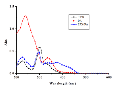
Figure 2 Absorption spectra of LFX [20µM], PA [20µM] and LFX-PA [20µM]in acetonitrile medium with slit width 1.0cm.
Absorption bands of LFX-PA (226nm and 298nm) got blue shifted by few nanometers from the absorption bands of LFX. The absorption ratio of band at 235nm of PA decreases sharply from 1.28a.u. to 0.316a.u., indicating 4 fold sharp absorption changes due to interaction of LFX with PA. A new broad band appeared in the absorption spectrum of LFX-PA at 375nm due to bright yellow color of the product formed.
Time for formation of LFX-PA ion pair: The effect of time on the formation of ion pair was studied carefully and illustrated in Figure 3. An absorption intensity of PA at 235nm was monitored over the reaction time. The sharp decrease of band maxima at 235nm indicated progress of the reaction in terms of ion-pair formation. It is clear from the Figure 3 that complete formation of ion pair needs 5-6 minutes before filtration.
Stoichiometry: The nature of binding of reagent to the drug was determined by continuous variation method.17 The results indicated that 1:1, [PA]: [drug] ion-pair is formed through the electrostatic interaction between positively protonated LFX and negatively charged PA, as shown in the proposed mechanism (Figure 4).
It is proposed in the mechanism that in the aqueous medium the reactants LFX and PA are more stable in their respective ionic forms and the two species are held together through electrostatic forces, resulting in the formation of ion-pair (LFX-PA). We suggested that compounds formed between LFX and PA have an ion-association character. This interpretation is in fair agreement with the results of the examination of fluoroquinolones compound and anionic forms of other organic substances.18-20
Effect of solvent: Levofloxacin is an amine containing two hetero atoms with lone pair as well as another nitrogen which is quaternary in nature and is devoid of any unshared basicity (basic character). The proposed method of detection of the antibiotic is based on its characteristics to form ion-pair with strongly acidic species like picric acid. The hypothesis that Levofloxacin forms stable ion pair has been verified by UV-Visible spectroscopy. UV-Vis spectrum of the substrate LFX, its ion-pair and the reagent PA were recorded separately in acetonitrile medium. The spectra were taken again by changing the solvent medium from pure acetonitrile to its mixture with 10% water and in pure aqueous media (Figure 5). A description of absorbance is as observed in all the three media are given in Table 1. Some interesting observations can be made from the absorbance values as summarized below:
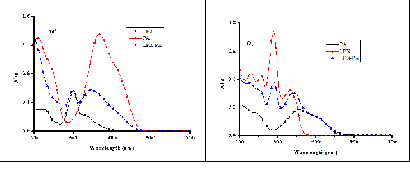
Figure 5 Absorption spectra of LFX [10-3M], PA [10-3M] and LFX-PA [10-3M] in (a) CH3CN:H2O medium (b) H2O medium (slit width 1.0cm).
|
S. no. |
Solvent |
Reagent |
lmax (nm) |
Absorbance Values |
|
1 |
CH3CN |
LFX [10-3M] |
226 |
0.277 |
|
PA[10-3M] |
235 |
1.28 |
||
|
LFX-PA[10-3M] |
220 |
0.349 |
||
|
335 |
0.238 |
|||
|
2 |
CH3CN:H2O (9:1; v/v) |
LFX[10-3M] |
301 |
0.568 |
|
PA[10-3M] |
209 |
1.305 |
||
|
LFX-PA[10-3M] |
295 |
0.547 |
||
|
3 |
H2O |
LFX[10-3M] |
228 |
0.439 |
|
289 |
0.738 |
|||
|
PA[10-3M] |
356 |
0.18 |
||
|
LFX-PA[10-3M] |
229 |
0.375 |
||
|
289 |
0.373 |
Table 1 Absorption values of LFX, PA, LFX-PA in different solvent systems
Effect of solvent was also studied on blank reagent i.e., picric acid which showed charge transfer absorption band at 330 nm in pure acetonitrile medium. This absorption band gets shifted to 366nm in case of CH3CN:H2O and 356nm with the band at 404nm in case of water as solvent system. These studies prove that water can be used as an excellent solvent for ion-pair formation.
Quantitative study:Picric acid has been used as a reagent for the spectrophotometric determination of Levofloxacin. Typical calibration graphs for the determination of the drug were obtained as shown in Figure 6 in different solvent systems. In case of acetonitrile, absorbance values for LFX-PA increased linearly with increasing concentration of LFX in the working range 6×10⁻⁵ - 24×10-5 M at 298nm, as shown in Figure 6a with correlation coefficient value 0.993, intercept 0.16965 and slope 0.02867. Similarly, calibration graphs were plotted for LFX in CH3CN:H2O (9:1; v/v) and H2O as solvent media in the concentration range (6×10⁻⁵-24×10-5M and 6×10⁻⁵-24×10-5M, respectively) which also showed linear responses with correlation coefficient values 0.966 and 0.967, respectively (Figure 6b and 6c).
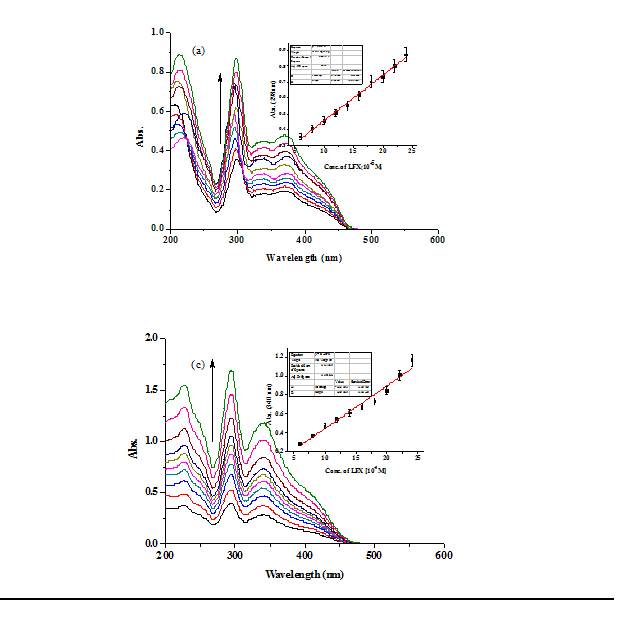
Figure 6 Absorption changes of LFX-PA with increasing concentration of LFX (6×10⁻⁵M-24×10-5M) in reaction mixture in (a) CH3CN (b) CH3CN: H2O (c) H2O as solvent medium with slit width 1.0cm. Insets showing their respective calibration plots.
Electrochemical determination of ion pair
Electrochemical behavior of LFX and PA:Cyclic voltammograms of LFX showed one major oxidation peak at 1.71 V with two minor oxidation peaks at 0.61V and 1.15V. The reduction peaks were observed at 1.65 V (major) and 1.13 V (minor) (Figure 7a), with their corresponding peak currents as shown in Table S2. The redox cycle at 1.7 V corresponds to quasi reversible process probably occurring at the piperazine moiety as observed from the peak current values.21 Differential pulse voltammetry also supported the observed peak potentials as shown in Figure 7b & 7c.
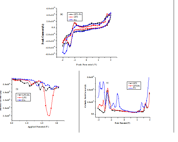
Figure 7 (a) Cyclic Voltammogram of LFX, PA and LFX-PA (5×10⁻⁴M) (b) Anodic Differential Pulse Voltammogram of LFX, PA and LFX-PA (c) Cathodic Differential Pulse Voltammogram of LFX, PA and LFX-PA in CH3CN containing 0.1M TBAPF6 as supporting electrolyte at scan rate 50mV/sec (Ag/Ag+ as a reference electrode).
|
S. No. |
Solvent |
Reagent |
Anodic Peak Potential (V) |
Anodic Peak Current |
Cathodic Peak Potential |
Cathodic Peak Current |
|
1 |
CH2CN |
LFX |
0.61 |
-2.54 |
1.73 |
0.13 |
|
PA |
1.33 |
-5.76 |
1.33 |
2.19 |
||
|
LFX-PA |
0.51 |
-2.73 |
1.69 |
4.52 |
||
|
2. |
CH2CN:H2O |
LFX |
-- |
-- |
-1.43 |
0.17 |
|
PA |
1.0 |
-27.23 |
-1.89 |
10.6 |
||
|
LFX-PA |
1.09 |
-24.10 |
-0.8 |
0.15 |
||
|
3. |
H2O |
LFX |
1.030 |
-22.18 |
-- |
-- |
|
PA |
1.55 |
-69.29 |
-0.98 |
45.02 |
||
|
LFX-PA |
1.56 |
-65.68 |
-0.49 |
14.44 |
Table S2 Peak potentials and corresponding peak currents of LFX PA and LFX-PA.
In case of PA, a redox cycle at 1.31V, cathodic peaks at 1.35V, -0.51V, -0.84V, -1.62V and -1.92V appeared which might be due to the reduction processes occurring at the nitro groups of PA. Likewise, two oxidation peaks at 1.63V and 1.82V were observed in the anodic cycle which corresponded a two step oxidation of phenolic hydroxyl group (Figure 7a). Similar results were observed in differential pulse voltammograms as shown in Figure 7b&7c.
Ion-pair determination voltammetrically: Ion-pair formed from the interaction of drug LFX and PA was studied for its peak potentials by cyclic and differential pulse voltammograms. As observed from the voltammograms (Figure 7b), anodic peak of LFX-PA (1.64V) got shifted by 70mV when compared with peak potential of LFX (1.71V) with large decrease in peak current from 25.4µA to 9.2µA. In case of LFX-PA, new peaks at 0.90V, 1.12V, 1.45V, 1.64V and 1.88V also appeared. Similarly, in case of cathodic cycle, peaks at 1.63V, -0.80V, -1.62V and -1.92V appeared which proved the interaction of the PA and LFX. Deviation in the peak potentials of LFX-PA from that of PA and large difference in peak current values might be due to the interaction of PA with the drug LFX.
Effect of solvent: Voltammetric determination of LFX-PA was also carried out in CH3CN:H2O (9:1; v/v) and H2O as solvent systems (Figure 8). It was observed that in CH3CN:H2O (9:1; v/v) there was a large increase in peak current value of anodic and cathodic peak potentials with small shift in their peak potentials. While in case of aqueous medium, oxidation occurs at a single peak potential (1.56V) with a large increase in the peak current i.e. 65µA. The results demonstrated the use of H2O as a perfect solvent because complete oxidation takes place at specific peak potential i.e., 1.56V having sufficiently high peak current as included in Table S2. Water being a highly polar solvent further increases the chance of oxidation occurring at the piperazine moiety and hence high peak current was observed as compared to CH3CN and CH3CN:H2O (9:1; v/v) as solvent media. In case of cathodic cycle of LFX-PA, it was observed that with increasing water content in solvent medium, cathodic peak potentials got shifted to lower values. These observations prove that oxidation of piperazine moiety and reduction of nitro groups can be easily carried out if we move from pure CH3CN to CH3CN:H2O mixture media increasing water ratio in solvent medium. Similar results were also observed in pure LFX and PA.
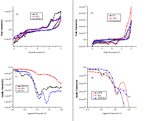
Figure 8 Voltammograms of LFX, PA and LFX-PA. (a), (c) and (e), in CH3CN:H2O. (b), (d) and (f), in H2O. (a) and (b), cyclic voltammograms. (c) and (d), anodic differential pulse voltammograms. (e) and (f), cathodic differential pulse voltammograms containing 0.1 M TBAPF6 as supporting electrolyte at scan rate 50 mV/sec. (Ag/Ag+ as a reference electrode).
Quantitative study Figure 9 shows the dependence of voltammetric peak current on Levofloxacin concentration value. It can be seen that peak current at 1.45V increases linearly with increasing LFX concentration from 15×10-4M – 60×10-4M with R2 = 0.994 with relative standard deviation 5% in CH3CN as solvent medium. Likewise, calibration graphs were plotted in CH3CN: H2O and H2O (Figure 9b &damp; 9c). From these calibration graphs, we can conclude that PA can be used as an anionic reagent for the determination of LFX in different solvent systems.
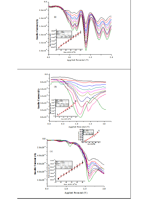
Figure 9 Differential Pulse Voltammograms of IP at different concentrations of LFX (15×10-4M - 60×10-4 M) in (a) CH3CN (b) CH3CN: H2O (c) H2O; Insets representating the calibration plots of their respective peak currents w.r.t. concentrations. (Ag/Ag+ as a reference electrode).
Computational study
Computational study of LFX, PA, LFXH+, PA-, LFX-PA- and LFXH+-PA- was carried out on Gaussian 03W software. All the structures were optimized for minimum energy by using DFT calculations. Basis sets used for optimization of structures and complexes were B3LYP/6-31G in gaseous state. The complex (LFXH+-PA-) was assigned net zero charge and all the optimizations were carried out in ground state. The DFT calculations of two types of complexes i.e. ionized and unionized gives us the energy of HOMO, LUMO, energy gap and gas phase energy of LFX, PA, LFXH+, PA-, LFX-PA- and LFXH+-PA- as shown in Table 2. Surfaces of HOMO’s and LUMO’s of LFX, PA, LFXH+, PA-, LFX-PA- and LFXH+-PA- appear in Figure 10. Energy gap of LFXH+-PA- gets reduced after ion-ion association complex and leads to the stability of the interaction.
|
Compounds |
Gas Phase Energy (eV) |
HOMO (eV) |
LUMO (eV) |
Energy Gap (eV) |
|
LFX |
-34355.629 |
-5.363 |
-1.430 |
-3.933 |
|
PA |
-25050.256 |
-8.613 |
-4.091 |
-4.522 |
|
LFXH+ |
-34366.540 |
-8.569 |
-4.105 |
-4.564 |
|
PA- |
-25037.086 |
-2.929 |
-0.148 |
-2.781 |
|
LFX-PA- |
-59407.980 |
-6.255 |
-3.035 |
-3.220 |
|
LFXH+-PA- |
-59393.749 |
-2.732 |
-0.303 |
-2.429 |
Table 2 Energy of HOMO, LUMO, gas phase energy and energy gap of various compounds and complexes.
The DFT calculations show that in case of LFXH+-PA-, picric acid lies perpendicular to the plane of LFX and appears as a V-shaped structure as shown in Figure 11. Hydrogen ion of picric acid gets ionized and interacts with nitrogen of piperazine moiety of LFX. Bond distance between terminal nitrogen of piperazine moiety and ionized hydrogen is 1.06A◦, while bond distance between phenolate oxygen of PA and ionized hydrogen gets elongated and is 1.62A◦ as measured from optimized geometry of the complex.
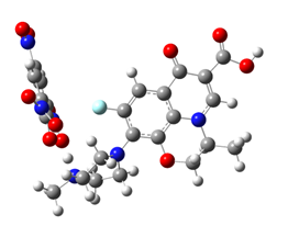
Figure 11 Interaction diagram of optimized structures of PA and LFX using DFT/B3LYP/6-21G method. Red balls represent oxygen, blue balls represent nitrogen and grey balls represent carbon atoms.
Analytical application
The proposed method was successfully applied for the determination of Levofloxacin content in commercial tablets. Commonly used additives in the manufacturing of tablets were not found to interfere in the analysis. To establish validity of this proposed method, recovery test was performed by using standard addition technique. Recovery is calculated as:
% recovery = (measured Levofloxacin ×100)/added Levofloxacin
Concentration values obtained by this method for different series of tablets are shown in Table 3. When the electrochemical procedure was applied, the anodic wave at 1.64V was monitored. Results given in Table 3 reveal that recoveries obtained were in the range 94.6% to 100% proving the validity of the proposed method.
|
Pharmaceutical Preparations |
Proposed Method |
|||||
|
Spectrophotometric Method |
Voltammetric Method |
|||||
|
Taken (mg/mL) |
Found (mg/mL) |
Recovery (%)* |
Taken (mg/mL) |
Found (mg/mL) |
Recovery (%)* |
|
|
Levomac tabletsa (Levofloxacin, 500 mg/tablet) |
20 |
18.9 |
94.6 |
20 |
19.3 |
96.7 |
|
Levac tabletsb (Levofloxacin, 750 mg/tablet) |
20 |
19.9 |
99.9 |
20 |
19.9 |
99.3 |
|
Levomac tabletsc (Levofloxacin, 250 mg/tablet) |
20 |
19.7 |
98.8 |
20 |
19.9 |
99.7 |
Table 3 Recovery studies using standard addition method.
*Average of at least 3 determinations.
aMacloeds Pharmaceutical LTD, Mumbai (Batch No. KLB502A).
bFranklin Laboratories Pvt. Ltd, Roorkee (Batch No. LYC75-14).
cMacloeds Pharmaceutical LTD, Mumbai (Batch No. ALC401C).
Spectrophotometric and Voltammetric methods were developed for the determination for Levofloxacin at ppm level. Picric acid having three nitro groups facilitates the removal of H+ ion from phenolic hydroxyl group, which eventually forms the ion association at the nitrogen center of piperazine moiety of Levofloxacin. The method described herein has many advantages; easy synthesis of ion pair complex, stability of the complex, less time consuming, sensitive, spectrophotometric as well as electrochemically active.
We are thankful to Saurav Chemicals, Derabasi (Patiala) and Dr. Manmohan Chhibber (Associate Professor, School of Chemistry and Biochemistry, Thapar University, Patiala) for providing the drug in pure form. Authors are also grateful to Director Thapar University, Patiala for providing research facilities.
Supplementary information includes Table S1 and S2 which gives us tabulated information of frequencies of LFX, PA, LFX-PA and Peak current values of LFX, PA, LFX-PA respectively. Supplementary Information is available at www.ias.ac.in/chemsci.

©2017 Mittal, et al. This is an open access article distributed under the terms of the, which permits unrestricted use, distribution, and build upon your work non-commercially.