International Journal of
eISSN: 2475-5559


Research Article Volume 1 Issue 4
1Atomic Minerals Directorate for Exploration and Research, India
1Atomic Minerals Directorate for Exploration and Research, India
2Department of Atomic Energy, India
2Department of Atomic Energy, India
Correspondence: Sukanta Goswami, Atomic Minerals Directorate for Exploration and Research, India, Tel 9492652413
Received: October 25, 2016 | Published: December 13, 2016
Citation: Goswami S, Mukherjee A, Zakaulla S, et al. Microbial mat related features in palaeoproterozoic gulcheru formation and their role in low grade uranium mineralisation. Int J Petrochem Sci Eng. 2016;1(4):106-112. DOI: 10.15406/ipcse.2016.01.00019
Interaction of microbial communities with clastic sedimentation is observed in Palaeoproterozoic Gulcheru Formation in Indian Purana stratigraphy. Both mat growth features and mat destruction features are preserved. Bacterial activity related to microbial mat formation is an important factor in producing a reducing environment. As compared to general uranium content in quartzite (0.5 ppm) and greywacke (3ppm) after Taylor in 1965, the Gulcheru Formation indicates higher average uranium content in grey quartzite (9.9 ppm, n=250), siltstone (15.42 ppm, n=500), pitted quartzite (5.6 ppm, n=400) and pink massive quartzite (7 ppm, n=800). It is also noted that the average uranium content is higher in and around the microbial mats which indicates the importance of mats in uranium entrapment. It is inferred that fertile fractured basement granitoid with average 40ppm U content is source rock, sandstone with reducing environment is host rock and ground water has acted as transporting agent. The siltstone and shale in the heterolithic facies at the top have played significant role in channelling uraniferous fluids to favourable sites and also at the same time prohibiting widespread flushing and dilution of fluids. In the context of Indian Purana stratigraphy and also of global Proterozoic biosphere, Gulcheru sandstone occupies a unique place as it preserves the influence of microbial communities on Palaeoproterozoic bio sedimentation.
Keywords: Microbial mat; Palaeoproterozoic; Gulcheru Formation; Cuddapah basin; Andhra Pradesh
Microbial mats are the earliest form of life on Earth for which there is good fossil evidence, from 3,500 million years ago, and have been the most important members and maintainers of the planet's ecosystems. These are laminated microbial communities that generally develop in aqueous environments under conditions that exclude fauna. The biogeochemical cycles in microbial mats are usually largely closed, although small fluxes of elements are exchanged with the geosphere, biosphere and atmosphere. Gulcheru quartzite hosts both disseminated and vein type of uranium mineralisation. In the sandstone-siltstone heterolithic facies, uranium is in adsorbed form with ferruginous material.1 Ever since the location of uranium mineralisation in the Cuddapah Basin,2 sustained exploration efforts with revised and improved geological concepts led to the discoveries of different types of uranium mineralisation in different lithologies and structures, in and around Cuddapah Basin.3 Microbial mats have increased the concentration of metal in many ore deposits, and without this it would not be feasible to mine them -examples include iron (both sulfide and oxide ores), uranium, copper, silver and gold deposits.4 In Indian Purana basins, reports of mat-induced sedimentary structures, preserved in siliciclastic rocks are mostly from the Mesoproterozoic sediments of Vindhyan basin5-8 and Cuddapah basin.9 Microbial activities and their evidences in Palaeoproterozoic siliciclastics are not yet reported from anywhere other than Gulcheru Formation.9
Studies with dissimilatory metal-reducing bacteria (DMRB) and dissimilatory sulfate-reducing bacteria (SRB) such as Desulfovibrio (D.) desulfuricans and D. vulgaris, which can use U(VI) as a sole electron acceptor, show that U(VI) is enzymatically reduced to U(IV) and removed from water.10-12 Mechanisms for the microbially mediated removal of U(VI) have been defined such as the surface sorption of U(VI) on cells, the abiotic reduction of U(VI) by SRB-produced H2S, and the enzymatic bioreduction of U(VI) with U(VI) acting as a terminal electron acceptor.13 The 3.5-billion-year survival of mats testifies to their capacity in adapting to and altering hostile environments through cellular and community-mediated activities.14,15 Stromatolites and associated carbonate structures and microbial mat features are frequently found in carbonate rocks due to mineral precipitating environments but sandy siliciclastic sub-aqueous environments are not typically characterised by mineral precipitation. However, uranium mineralisation in sandstone occurs in presence of carbon and/or pyrite, which can act as reducing agents. An important feature of microbial mats is their carbon autotrophy, i.e., the fixation of inorganic carbon.
The present paper reports the probable influence of microbial communities on low grade uranium mineralisation and an elaborative descriptions of different mat related mesoscopic features in Gulcheru quartzite for the first time.
The Cuddapah Basin Figure 1 occupies an area of about 44,500 sq km on the eastern part of the Dharwar Craton in the Indian shield and consists of sedimentary and associated volcanic rocks of about 12 km thickness, ranging in age from late Paleoproterozoic to Neoproterozoic.16-18 Lithostratigraphically, the Cuddapah Basin is divided into Papaghni, Chitravati, Nallamalai and Kurnool groups from base to top. A detail description of stratigraphy, structure, igneous activities, mineral potentiality and basin evolution is given by Nagaraja Rao et al.19 The Cuddapah Supergroup is composed dominantly of argillaceous and arenaceous sequences with subordinate calcareous sediments deposited in peritidal complex with shallow marine carbonate shelf and beach.19 The lowermost Palaeoproterozoic Papaghani Group, with basal polymictic conglomerate, siliciclastic-dominant Gulcheru Quartzite and overlying mixed siliciclastic-carbonate bearing Vempalle Formation, unconformably overlies the Archaean Dharwar batholithic granitoids with remnants of greenstone belts. The present study area Figure 2 along south-western margin of Cuddapah Basin forms part of Papaghni Group with basal Gulcheru Quartzite. The general strike of the formations in the study area (78˚20′E to 78˚35′E, 14˚15′ N to 14˚20′N, Toposheet Nos. 57 J/7 and J/11) is E-W, dip ranges from 15˚ to 25˚ due N.
The Papaghni sub-basin in the western part of the Cuddapah Basin hosts the Palaeoproterozoic rocks whose age of sedimentation has been well constrained.16,20 Based on the study of several outcrops in this area, a composite lithology showing lithofacies stratigraphically from base to top has been prepared Figure 3. The basal Gulcheru Quartzite consists of a matrix to clast-supported thick-bedded polymictic conglomerate with occasional interbeds of gritty and trough cross-bedded feldspathic sandstone. Subangular-subrounded pebbles of vein quartz, pegmatite, granite, fine micaceous sandstone, black chert and grey shale/argillite suggest their derivation from the granitoid basement with greenstone patches. The coarse gritty matrix consists of quartz and pink feldspar, locally with ferruginous patches. The common occurrence of trough cross-bedding in the gritty interbeds, channel lags, outsized clasts, lateral thinning out of the stack of conglomeratic beds and a generally fining-upwards facies suggest an alluvial-fan setting21 for the lowermost unit. The ~20m thick basal conglomerate - gritty feldspathic sandstone facies grades upward to massive quartzite with brick red coloured Fe-rich bands. This facies of ~15m thickness indicates rapid deposition from high concentration sandy suspension flows. The particles settling from suspension are prevented from being reworked after reaching the depositional surface due to quick burial. This normal graded sandstone indicates deposition from sandy suspension flows by gradual failure of capacity and competence and hence, particle size decreases upward. Higher up in the section massive ferruginous quartzite units are overlain by medium to fine grained grey quartzite, with occasional rippled to cross stratified arenitic interbeds (20m). This unit consists of well sorted sub-rounded quartz sand with rare feldspar grains. The overlying greyish yellow coloured well rounded medium to coarse grained multilayered sandstone (5m) exhibit presence of trough cross stratification with individual set thickness varying from 10cm to 50cm. This is followed by multilayered rippled sandstone facies (15m), which is characterised by the presence of different types of ripples like rhomboid ripples, flat topped ripples, double crested ripples and different types of interference ripples like ladder back ripples. Heterolithic facies (12m) of alternative siltstone and sandstone comprises purple to grey coloured siltstone, silty sandstone and sandy siltstone and sandstone layers. The overlying pitted quartzite (~5m) shows circular pits developed due to interplay of different physico-chemical factors which prevailed in syn to post depositional environments.22 In addition to different agents of physical weathering which played a vital role in the formation of pits in the Gulcheru Quartzite, syn-depositional precipitation of iron oxides and syn to post diagenetic chemical changes are believed to be the main factors.22 The topmost unit crops out near the contact of Vempalle Dolostone and consists of heterolithic dark brown micaceous iron rich reddish shale - fine sandstone (14 m) with cross strata, mudcracks and occasional lag pebbles. The association of straight-crested, interference and flat-crested ripples, shallow troughs, bipolar current azimuths, mudcracks in the heterolithic facies and occasional lag pebbles suggest the uppermost part of the Gulcheru Quartzite to be of tidal-flat origin. Therefore, a transgressive sequence can be seen in Gulcheru Formation. Aquatic deposits are constantly reworked by water motion, and therefore all benthic organisms must be able to tolerate the physical sediment dynamics caused by waves and currents.23 The photoautotrophic cyanobacteria, and many other microbial mat-forming bacterial groups are highly mobile organisms, and can move actively through the sediment.24 Therefore, in addition to the primary sedimentary structures the microbial mat related structures have also been studied carefully.
In response to erosive forces, filamentous microorganisms stabilize the sediment by entangling the mineral particles like an organic meshwork, and the adhesive mucilages called, extracellular polymeric substances (EPS), glue the mineral particles together.25 This two-fold microbial sediment fixation is termed bio stabilization,26 which encompasses binding, trapping and baffling of clastic sediments. Thin bio films comprising intermingled sand grains and microbial filaments tend to stabilise sediment surfaces following physical reworking processes. If sedimentary particles are deposited on the sea floor, the microorganisms move upward to keep up with the rising sedimentary surface, simultaneously accumulating sediment by baffling and trapping. However, during periods of calm hydraulic conditions, the microbial cells assemble in the sedimentary surface layer to form the organic meshwork of a carpet-like microbial mat. Those activities of benthic prokaryotes in response to the sediment dynamics form characteristic ‘microbially induced sedimentary structures (MISS).27-30
In the present context a broad description of mat related features is necessary since the Gulcheru Formation occupies a unique place as it preserves the influence of microbial communities on Palaeoproterozoic bio sedimentation9 and reports for the first time about the presence of different categories of MISS. The MISS of the studied area are classified into two types, i.e., mat growth features and mat destruction features.
Palimpsest ripples and loaded ripples
The loaded ripple shows rippled sandstone surface marked by loading features related to a thin veneer of overlying sandstone Figure 4A. The evidence for soft sediment deformation of the lower sandy bed and the lack of amalgamation of the two sandy layers is indicative of the presence of mat to provide flexibility and cohesiveness of the lower deformed bed.31 Similarly, pelimpsest ripples32 in sandy sediments are preserved with crests being relatively sharp and un-reworked. Absence of mud between successive rippled sandy beds is typical character to identify Figure 4B. These are indicating intertidal marine conditions.31
Wrinkle structures
These are found either as bed tops or bed soles showing a reticulate pattern of small raised ridges Figure 4C and reflects original topography of a microbial mat growing on sandy substrate, or such microbially bound sediment affected by micro scale loading or shear stresses. Wrinkle structures are usually associated with ‘kinneyia’ and indicates subtidal to intertidal environment. These are thought to form by either purely microbial processes or a combination of these with bio film mediated physical loading processes.
Exfoliating sand laminae
Successive, very thin (≤1mm) veneers/layers are found to be removed Figure 4D. The lack of any mud separating sandstone veneers as well as variable OES textures on each of the underlying layers suggest that each thin sandy layer was resistant to erosion, loading and overprinting by the succeeding thin sand veneer due to the presence of bio film upon each sandy layer. These are usually indicating intertidal to subtidal marine conditions.

Patchy ripples
Flat sandstone surfaces in which patches are marked by ripples of various types, and where the ripples grade into the surrounding sandstone surfaces Figure 4E & 4F are indicative of intertidal environment in the study area. These are formed due to current and wave actions in patches within a mat stabilised sandstone bed surface where the mat is either absent or has been removed by reworking processes.31
Apart from this presence of kinneyia ripples and old elepant skin (OES) structures are also reported earlier by Chakrabarti & Shome.9 The kinneyia ripples show irregular mosaic-like pattern are occasionally associated with wrinkle marks in the heterolithic facies of the Gulcheru Sandstone in the study area. Walls of pits or grooves are steep sided and occur on sandstone or muddy sandstone bed tops or, flat tops between truncated ripple crests. These are formed due to build-up of gas bubbles in sandy sediments immediately underlying a bio film or mat. Old elepant skin (OES) structures in multilayered rippled sandstone facies shows quasi-polygonal textures. Bed top OES bears concave bulbous depressions and sharp convex crests. Bed bottom OES bears convex to bulbous textures with sharp concave depressions. These are usually associated with oscillatory ripples and exfoliating sand laminae in intertidal to subtidal marine conditions.31
Sand cracks
These are isolated spindle shaped cracks formed by tearing of mat. The triple junction or tri radiate intersections of incipient cracks Figure 5A may turn into complex intersection of either essentially straight cracks or less commonly, curved crack patterns. Finally, there appears to be a second generation of more complex curve cracks with smaller widths Figure 5B, which show no relationship to triple junction features. All these crack features are inferred to reflect mat desiccation and sub aerial shrinkage, with concomitant cohesiveness provided by the mat to the underlying sands which then preserve the same features.33
Curled crack margins
These are mat surface with shrinkage cracks in which crack margins show curling. In rock record irregular sub circular Figure 5C or linear ridges Figure 5D occur which are distinctly flattened and may be inferred to reflect the curled margins of sub-circular openings in thin microbial mat that previously covered a sandy sediment surface or a narrow linear crack with curling edges.33 These form subaerially that undergoes shrinkage and cracking in upper intertidal to lower supratidal conditions.
Filled sand cracks
These resemble sand cracks but in contrary to that these are positive ridges. The cracks were filled from above by other sands brought in either by aqueous currents or aeolian actions.33 On the decay or erosion of the mat, these crack fills form positive ridges,32 with the similar plan-form geometry as the original microbial shrinkage cracks Figure 5E & 5F.
Petee ridges
The sandstone making up the petee ridges are composed of the same material as that comprising the containing bed Figure 5G. These are representative of braid deltaic-tidally controlled epiric marine coast line.33
Manchuriophycus or Rhysonetron
In the upper rippled (or interference rippled) surface of sandstone beds, within ripple troughs, complex crack patterns with curved, circular, spiral, sinuous or tri-radiate forms occur, which follow the edges of these troughs Figure 5H. These are indicator of coastal plains of lacustrine or shallow marine environments and may be associated with significant tidal actions.33
Uranium mineralisation and microbial mat
Organic matter has long been known to be associated with many types of mineral deposits, especially certain types of uranium deposits. Such an association of uranium and carbonaceous material can be attributed to metal organic compounds or to adsorption and precipitation mechanisms during the sedimentation.34 There are a few Precambrian sandstone hosted uranium deposits associated with carbonaceous matter of probable algal origin.35 Uranium concentration in siliciclastic sediments is governed by its detritus source and the compositional and textural properties. As compared to general uranium abundance in quartzite (0.5ppm) and greywacke (3ppm),36 the Gulcheru Formation indicates higher average uranium content in grey quartzite (9.9 ppm, n=250), siltstone (15.42 ppm, n=500), pitted quartzite (5.6 ppm, n=400) and pink massive quartzite (7ppm, n=800).1 In the study area, several spotty uranium anomalies have been noted with radiometric assay values ranging up to 0.034% U3O8. However, along the E-W trending faults in Madyalabodu area higher grade mineralisation has also been recorded intermittently over a strike length of 1600m in Gulcheru quartzite showing 0.0207% eU3O8 to 0.162% eU3O8.37

Apart from the fracture controlled mineralisation, the Gulcheru sandstone also hosts uranium deposited at oxidation – reduction interfaces where presence of carbon and/or pyrite might have acted as reducing agents. This is further corroborated by the development of microbial mat in these Palaeoproterozoic sediments. An important feature of microbial mats is their carbon autotrophy in which the fixation of inorganic carbon is a key process. Besides, oxygenated ground water has played a significant role leading to leaching of uranium. Local reducing conditions were swamped by oxidising ground water and thus dark sandstone have become reddish through the in situ oxidation of ferrous minerals to ferric oxyhydroxide, a precursor of hematite Figure 6. Pitchblende and coffinite are reported as dominant uranium minerals in anomalous zones.1

The feldspar component of the host Gulcheru rocks indicate a granitic source while an environment of rapid erosion and sedimentation have provided the required hydro-physical conditions, particularly permeability for adequate groundwater migration. Impermeable or less permeable siltstone and shale in the heterolithic facies and at the top have played significant role in channelling uraniferous fluids to favourable sites and also at the same time prohibiting widespread flushing and dilution of fluids. The studied area consists of numerous microbially induced sedimentary structures, indicating presence of carbonaceous organic matter which may be destroyed at places closer to the anomalous zones by oxidation. However, the oxidation potential of ground water gradually decreases and reaches to localised reducing environment, which leads to the precipitation of uranium from solution. This is also apparent from precipitation of pitchblende or coffinite with organic debris exhibiting syngenetic sedimentation pattern.38
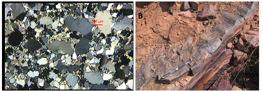
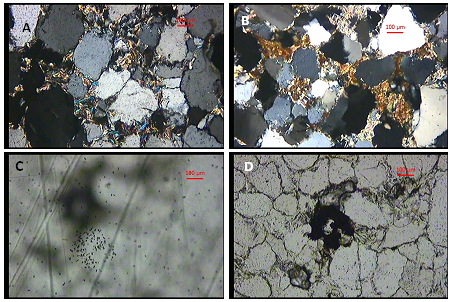
A large number of microbial mat-induced structures are reported from Mesoproterozoic and Neoproterozoic5,39-41 and Archean28 rocks. The record of global oxygen build-up also suggests proliferation of microbiota during the Mesoproterozoic.39-41 In the context of Indian Purana stratigraphy and also of global Proterozoic biosphere, it had been suggested by Chakrabarti and Shome 2010 that the Gulcheru sandstone occupies a unique place as it preserves the influence of microbial communities on Palaeoproterozoic bio sedimentation. Under microscope, mat related features can be understood from Figure 7A. Low order radioactivity recorded around mat and wave rippled sandstone Figure 7B near Madyalabodu area points to the significance of microbial activity in entrapment of uranium by adsorption and/or precipitation by reduction as depicted in the model Figure 8. The quartz grains are bounded by micaceous minerals indicating existence of mat and siliciclastic minerals were surrounded by an organic matrix. The gradual decay of organic matrix left behind the mica and clay minerals. The interstitial spaces of quartz grains are occupied by micaceous minerals Figure 9A and iron oxide in the form of limonite and goethite Figure 9B related to microbial mat which might had bind the detrital grains and provided reducing conditions for fixation of uranium. The rock samples from the mat bearing units have provided sparse to low density alpha tracks after 4days of exposure of CN film Figure 9C & 9D. The source of radioactivity is adsorbed uranium on iron oxides along interstitial spaces which is the most striking features to make out about the role of mats in uranium mineralization.


Based on detailed geological and radiometric studies of Gulcheru sediments and in depth studies on microbially induced sedimentary structures the following conclusions are drawn.
Impermeable or less permeable siltstone and shale in the heterolithic facies and at the top have played significant role in channelling uraniferous fluids to favourable sites and also at the same time prohibiting widespread flushing and dilution of fluids. Gulcheru Formation occupies a unique place in Cuddapah stratigraphy as it preserves the low grade microbially influenced uranium mineralisation in Palaeoproterozoic rock sequence.
We express our sincere gratitude to Shri P.S.Parihar, Ex Director, AMD for encouragement and support. The in charge petrology lab is thankfully acknowledged.
The author declares no conflict of interest.

©2016 Goswami, et al. This is an open access article distributed under the terms of the, which permits unrestricted use, distribution, and build upon your work non-commercially.