International Journal of
eISSN: 2574-9889


Letter to Editor Volume 1 Issue 1
1FRIGE Institute of Human Genetics, India
2Jena University Hospital, Institute of Human Genetics, Germany
Correspondence: Frenny J Sheth, FRIGEs Institute of Human Genetics, FRIGE House, Jodhpur Gam Road, Satellite, Ahmedabad-380015,Gujarat, India, Tel 91-79-26921414, Fax 91-79-26921415
Received: December 09, 2016 | Published: December 15, 2016
Citation: Sheth FJ, Naznin L, Liehr T, et al. FISH – the best technique in characterization of prenatally detected small supernumerary marker chromosomes (SSMC). Int J Pregn & Chi Birth. 2016;1(1):19-20. DOI: 10.15406/ipcb.2016.01.00005
small supernumerary marker chromosome, fish, karyotyping, prenatal
sSMC, small supernumerary marker chromosomes; FISH, fluorescence in situ hybridization; aCGH, array comparative genomic hybridization
Small supernumerary marker chromosomes (sSMCs) comprise additional genetic material of unknown origin identifiable by banding cytogenetics. Their frequency in newborn is 0.044%, in prenatal 0.075%, in mentally retarded 0.288%, and in subfertile cases 0.125%.1 In 70% of the cases, the sSMC is de novo, in 20% is inherited from the mother, and in 10% is inherited from the father. To mention, 70% of the sSMC carriers are clinically normal.2 The phenotypic expression of sSMCs is variable, from asymptomatic to symptomatic depending on their size, shape, chromosomal origin, satellite/non-satellite, genetic content i.e. euchromatic/heterochromatic and the level of the mosaicism; moreover uniparental disomy of sSMC’s sister chromosomes may also influence the clinical outcome.2,3
sSMCs are a major clinical problem, especially when detected prenatally during cytogenetic analysis. The risk for an abnormal phenotype in prenatally ascertained de novo cases with sSMC is ~13%. This has been refined to 7% for sSMC from acrocentric autosomes (excluding 15s) compared with ∼28% for non-acrocentric autosomes4 and 30% for all sSMC carriers.1 Strikingly, 30-50% of pregnancies diagnosed with sSMC go for termination.1 Since there is great variation in the clinical outcome, characterization of prenatally detected de novo sSMC is of significant importance to offer appropriate medical care and genetic counseling for the parents.
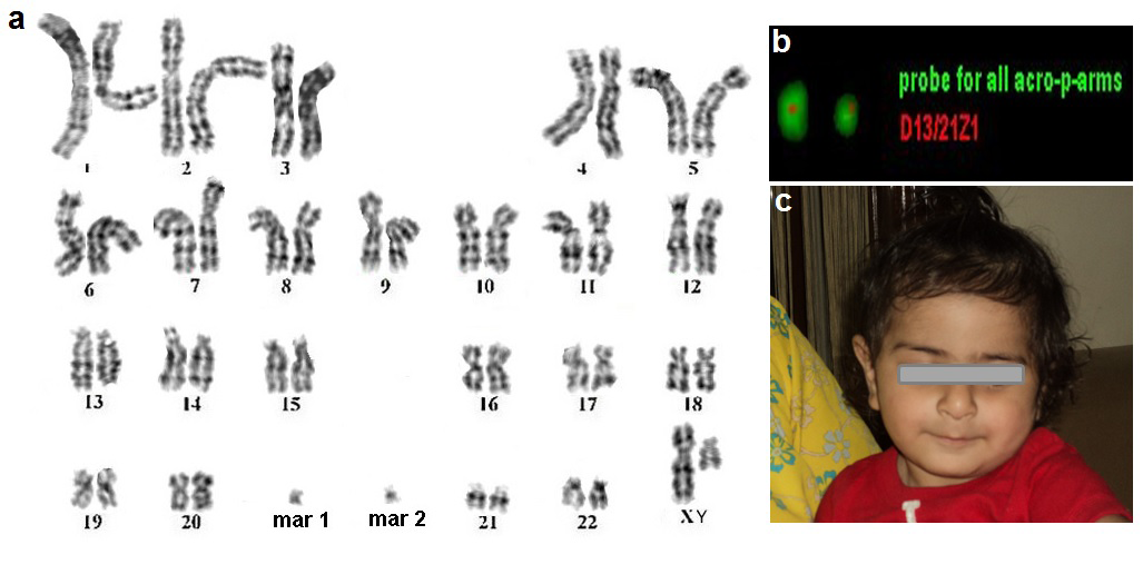
In the present case, prenatal study was advised to an elderly primi gravida due to advanced maternal age. Karyotypic analysis of the fetus carried out from the amniocytes showed presence of two sSMCs (i.e. 48,XN,+mar1,+mar2)[100%] with normal gonosomes (Figure 1A). Parental chromosomal analyses at 500 band resolution were normal, confirming de novo origin of both the SMCs. Triple marker study carried out at 16weeks did not show any risk for trisomy 13, 18 and 21. Fetal anomaly scan carried out at 16 and 24weeks were also normal. Various Fluorescence In Situ Hybridization (FISH) probes like (acro-)cenM-FISH, besides immunohistochemistry using antibodies CENP-B (all centromeres apart from Y) and CENP-C (all active centromere) characterized the sSMCs as inv dup(13 or 21)(q10) and del(13 or 21)(q10) (Figure 1B). Hence, the karyotype resulted to be 48,XY,+inv dup(13 or 21)(q10),+del(13 or 21)(q10)dn. According to FISH, the sSMCs most likely were derived from each other and consisted exclusively of heterochromatic material causing no clinical abnormalities. Hence, pregnancy was continued and delivered a normal baby boy and no dysmorphism was detected after birth (Figure-1C).

Current molecular cytogenetic technologies cenM-FISH approach, microdissection and reverse painting appearing to be the most straightforward way to determine the origin of sSMC. SubcenM-FISH approach is for characterization of the content and structure of the sSMC. Array Comparative Genomic Hybridization (aCGH) encompasses only euchromatic region and may be non-informative in such cases due to a lack of coding regions. Thus, FISH appears to be the best technique for the characterization of the mysterious chromosome presented as markers and helpful to define the genotype-phenotype correlations associated with sSMC. In developing countries, with very few laboratories being equipped with such complex molecular cytogenetic techniques, it is vital to address the issue that the origin of sSMCs in an abnormal karyotype needs to be studied in detail before advising termination. The wise use of such advanced cytogenetic techniques is clinically important to save the healthy fetus from getting aborted and also aid in genetic counselling for subsequent pregnancy.
We thank Dr. Krati Shah and Dr. Sunil Trivedi for giving critical comments. We also thank the family for their kind cooperation. The case report under study has not received any funding for any purpose.
The authors declare that they have no financial or non-financial competing interests.

©2016 Sheth, et al. This is an open access article distributed under the terms of the, which permits unrestricted use, distribution, and build upon your work non-commercially.