International Journal of
eISSN: 2574-8084


Literature Review Volume 6 Issue 1
Instituto Nacional de Cancerología, Colombia
Correspondence: Humberto Martínez-Cordero, Unidad de Hemato-oncología Instituto Nacional de Cancerología Servicio de Hematopatología Instituto Nacional de Cancerología, Colombia
Received: August 30, 2018 | Published: February 13, 2019
Citation: Martínez-Cordero H, Olivera LE, Escobar BP, et al. Langerhans cell histiocytosis in adults: report of five cases and literature review. Int J Radiol Radiat Ther. 2019;6(1):28-32. DOI: 10.15406/ijrrt.2019.06.00208
Langerhans cell histiocytosis (LCH) is a rare entity of unknown origin, currently considered as an orphan disease. Multisystem involvement is frequent. The disease is commonly associated with central diabetes insipidus (CDI) and other endocrinopathies such as hypothyroidism and type 2 diabetes mellitus. Few cases have been reported in children and the incidence rate in adults is estimated at one case per million. LCH may cause single-system or multisystem involvement, with bone and lung as the most commonly affected organs. The BRAF V600E mutation is present in more than half of the cases and is strongly associated with the presence of CDI. Currently, there are no definite treatment guidelines for adults. Cases of pulmonary involvement may respond to smoking cessation, although local resection or systemic chemotherapy may be required. LCH shows a good response to regimens based on vinblastine (VB) and prednisone (PDN). Here we report five adult cases diagnosed and treated at the National Institute of Cancerology in Colombia between 1997 and 2017. At some time point, all of the patients had multisystem disease, mainly bone involvement; four showed an associated endocrine disorder (particularly CDI), and in two patients the genital tract was affected. Three patients developed a metachronous cancer. First-line treatments were based on etoposide, VB, and PDN; response was good. Cladribine (2-CdA) was used as a rescue therapy in four cases reaching a global response rate of 100%; response was complete in four cases. Excellent response to the rescue regimen with 2-CdA suggests this might be an excellent first-line treatment strategy.
Keywords: langerhans cell histiocytosis, cladribine, BRAF
Langerhans cell histiocytosis (LCH) is a clonal disorder of unknown origin and variable clinical course, resulting from the abnormal proliferation of the myeloid dendritic cell line. Only 1-2 cases per million are identified in adults.1,2 Despite some familial cases have been reported, there is no known genetic susceptibility.3 LCH belongs to the L group of the Histiocyte Society classification, along with intermediate cell histiocytosis, Erdheim-Chester disease, and mixed disease.4 The BRAF V600E mutation is present in 55% of the cases; since it is more frequently found in multisystem presentation, it seems to be an important risk factor for resistance to treatment based on vinblastine (VB) and corticosteroids.5 In children, the disease has been described with a higher prevalence between one and three years of age, while in adults the issue is poorly defined.6 LCH can affect any tissue; nonetheless, involvement of heart and kidney is extremely rare.7
LCH was first described by Alfred Hand in 1893, when he reported the case of a 3-year-old boy with exophthalmos, polyuria, and localized rickets with skull lesions. In 1921, Hand described six additional cases with similar characteristics.8,9
Three diseases described in the past (i.e., Hand Schuller Christian disease, Letterer-Siwe disease, and eosinophilic granuloma), each with a different prognosis, should be currently classified as LCH; thus, avoiding such terms is highly recommended.10 The first classification of histiocytosis was published in 1987 by the Histiocyte Society. It included three categories: LCH, non-LCH, and malignant histiocytosis.11 In 2016, a new five-group classification was developed and LCH was placed in group L.12
At present, patients may be classified as suffering single-system or multisystem disease. The former may be either unifocal or multifocal (e.g., several bones involvement). In multisystem disease, two or more organs are involved including or not risk organs (e.g., the hematopoietic system, liver, and spleen), which may lead to an adverse prognosis.13 Involvement of the skull, especially bones of the orbit, as well as sphenoid, ethmoid, and temporal bones increases the risk of central nervous system (CNS) disease; therefore, those bones should also be considered as risk organs.14 Lungs were formerly considered as such; however, pulmonary disease may be improved by quitting smoking and lung involvement is not currently deemed as conferring an adverse prognosis.15 Consequently, some authors have considered pulmonary LCH should be left out of the risk group because of its different and well-defined etiology and prognosis.16
Histologically, LCH is characterized by phagocytic mononuclear cells proliferation with expression of CD1a and S100 proteins; Birbeck granules may be seen by electron microscopy. However, the latter has been replaced by CD1a and CD207 identification by immunohistochemistry.17 Strong associations with central diabetes insipidus (CDI) and secondary neoplasms have been described, but there are few reports of a clear association of LCH with endocrinopathies such as hypothyroidism and type 2 diabetes mellitus (DM).18 There are no accepted international guidelines for diagnosis and treatment specifically in adults. Most recommendations reflect experts consensus opinion based on the best evidence available.19
Summarizing treatment recommendations, quitting smoking and defining the best local treatment is indicated in single system disease, while in multisystem disease single-agent chemotherapy or combined chemotherapy are preferred.20,21
The clinical records of patients diagnosed and treated at the Department of Hematology of Colombia´s National Cancer Institute between 2001-2017 were analyzed. Five patients (three females) with a mean age of 58.6 years at the time of diagnosis were included in our series (Table 1). None of them reported smoking. Two patients presented with single system disease; nevertheless, at some time point all of the patients showed multisystemic involvement. Bone was the most frequently affected tissue; the skull was involved in four cases. No patient showed bone marrow involvement. Lungs and skin were affected in two patients. Strikingly, two of the three females showed genital tract involvement. Four patients had endocrinopathies, of which diabetes insipidus was the most frequent one; DM and hypothyroidism were also common. Three patients had metachronous malignancies.
|
Case |
1 |
2 |
3 |
4 |
5 |
|
Gender/age |
F/63 |
M/60 |
M/61 |
F/54 |
F/55 |
|
date of diagnosis |
2001 |
2001 |
2011 |
1997 |
2017 |
|
Immunopheno type |
S100+, CD1a+. Ki67 70%. PLAP- |
Strong expression of S100; CD1a. Few mixed cells CD68+. Proliferation index of 80% approximately (biopsy was taken at the time of relapse) |
S100+ and CD1a+. p63, CD56, AE1, AE3, CD20, NSE, HMB45, and sinaptofisin were all negative |
S100+. Lysozyme, ACL, CD20, keratin, EMA, chromogranin, and antimuscle factor were all negative |
CD1a+, S100+, CD68+, vimentin+, HLA-DR+. CD34, CD117, MPO, Cd30, and ACL were all negative |
|
Involvement |
Single-system disease. Genital involvement. A second relapse including CDI was documented eleven years later. A jaw lesion was documented at the last assessment. No tumor was reported |
Multisystem disease. Initial lung involvement; progression on skin (scalp, right axilla, groins) was documented in 2005 |
Multisystem disease. Bone (bilateral paranasal sinuses, orbital bones, long bones) and anorectal region involvement. No lung involvement was documented |
Single system involvement. Multisystem disease at the time of relapse. Cervix involvement; resection and RT followed. Subsequently, involvement of the vaginal cupola was documented. No lung or bone involvement |
Multisystem disease (skin, thoracic soft tissue, vertebral bone, right axillary adenopathy, a costal arch lesion spreading to pleura and thyroid) |
|
Treatment description |
CP + PDN: CR. Relapsed two years later (2003). ETO + VB were given as rescue regimen. Following second relapse, 2-CdA (five cycles) was given through August 2015 |
ETO + PDN. VB followed by MP + PDN and ETO as maintenance therapy every three weeks: PR. Progression was reported afterwards. 2-CdA (five cycles) was prescribed: PR |
VB + PDN (six cycles) with no response. Persistent bone involvement |
Hysterectomy with pelvic lymphadenectomy + brachytherapy. Vaginal cupola relapse (2007) treated with RT, PDN and VB: CR. The second relapse occurred with base skull bone, spine (C3), and auditive nerve involvement (severe hypoacusia) |
2-CdA as first-line therapy |
|
Cladribine therapy |
Five cycles through August 2015 |
Five cycles through August 2010; subsequently, zoledronic acid |
Four cycles ending on November 2013 |
One cycle. September 2009 |
Five cycles |
|
Outcome |
CR. Hypermetabolic jaw lesion documented by PET/CT; however, a biopsy showed few reactive lymphocytes (CD3) and plasma cells (CD38). Ki67 2%. CD20-, CD1a-, CD68-, S100-, CD56- |
PR. Improvement of cutaneous involvement |
CR confirmed by PET/CT |
CR confirmed by PET/CT |
CR confirmed by PET/CT |
|
Secondary malignancies |
NR |
Low-risk basal cell carcinoma (2011) |
Papillary thyroid carcinoma metastatic to lung |
Gastric adenocarcinoma (linitis plastica). Studies were negative for histiocytosis |
NR |
|
Other diseases |
Hypothyroidism. Hypopituitarism? |
Corticosteroid-induced DM. CKF |
Hypothyroidism. Insulin-requiring type 2 DM. HBP. CKF |
No |
Hypothyroidism. Insulin-requiring type 2 DM |
|
Patient status |
Alive without relapse |
Dead. No relapse at the time of last follow-up (2014) |
Last follow-up in |
No relapse at the of last follow-up (2016) |
Alive; CR |
Table 1 Data of the patients included in the series
2-CdA, cladribine; CKF, chronic kidney failure; CP, cyclophosphamide; CR, complete response; DM, diabetes mellitus; ETO, etoposide; HBP, high blood pressure; IMRT, intensity-modulated radiotherapy; MP, mercaptopurine; NR, not reported; PDN, prednisone; PET/CT, positron emission computed tomography; PR, partial response; RT, radiotherapy; VB, vinblastine
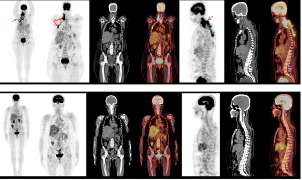
Most frequent first-line treatments were prednisone (PDN) and VB (three patients), etoposide (ETO; two patients), cyclophosphamide (CP), and radiotherapy (RT) (one patient each). Only one patient received maintenance therapy with mercaptopurine (MP), PDN, and ETO (Figures 1–3).

Figure 1 Study of F (18) –FDG PET-CT basal (above) and six months after treatment (below). From left to right: MIP (maximum intensity projection), coronal sections (PET, CT and fusion) and sagittal sections (PET, CT and fusion): Infiltrative hypermetabolic lesion involving the soft tissues of the dorsal region of the neck and of the right hemithorax, with extension to the posterior costal arches, the intersostal and the ipsilateral pleura (red arrow). Right axillary hypermetabolic adenomegaly (green arrow). Six months later there is a complete metabolic response.

Figure 2 40X, HE. Histocytic cells with nuclear clefts accompanied by abundant mixed inflammatory infiltrate with numerous eosinophils.
Twenty cycles of 2-CdA were given to five patients as a rescue regimen, with an average of five cycles per patient. One patient received the same schedule as frontline therapy. No associated toxicities were identified. Assessment of treatment response was possible in all cases. Overall response rate was 100%, with four complete responses and one partial response. Four patients were alive at the time of the last follow up, which is still ongoing. Only one patient died of a cause not related to a LCH relapse.
To the best of our knowledge, this is one of the largest series reported in Latin America.22 A total of 274 HCL cases in adults from 13 countries were registered between 2000 and 2001 with the International Rare Histiocytic Disorders Registry (IRHDR) of the Histiocyte Society. At present, we would be registering at the IRDHRD more patients than the United Kingdom, Australia, Greece, Switzerland or Slovenia.6
We highlight the fact that two female patients had genital involvement, a situation that had been previously reported in the literature. Both of them relapsed with multisystem disease. One female showed the same initial involvement in addition to CDI; the other one relapsed with involvement of cranial, vertebral, and ear bones, which led to severe deafness.23 Finding no reports describing initial single system disease followed by multisystem relapse is noteworthy.
The most frequent regimens used as front-line therapies were based on steroids plus VB, ETO, and RT. Based on the description made by Adam et al.,24 who reported a low toxicity, very good tolerance, and excellent durable responses, 2-CdA was used as front-line therapy in one patient.24 Four patients received 2-CdA as a rescue therapy (average of five cycles per patient). No relapse has been documented until today. There was just one case of soft tissue infection related to 2-CdA as the only adverse event.1,25
Another interesting finding was the association of LCH with second malignancies such as basal cell carcinoma, papillary thyroid carcinoma, and gastric cancer (linitis plastica). A high association with secondary malignancies in adults had been previously reported.26 We also found a strong relationship with hypothyroidism and type 2 DM as described in case reports, small series of patients, and the IRHDR.3 The limitations of our study are its retrospective nature and the lack of complete diagnostic tests, particularly molecular studies.
LCH is an extremely rare disease of clonal origin and an indolent course in most described cases, manifesting mainly on the skeletal system. Presence of the BRAFV600E mutation at least in half of the patients would be the reason for resistance to VB regimens. Pulmonary disease is related to smoking habit and shows a good response to cigarette cessation. Therapy is poorly defined and is based on VB, ETO, and steroid combinations. Given the promising disease control results, 2-CdA regimens seem to be one of the best treatment options. Cladribine has a great safety profile even as a front-line therapy with prolonged results. We hope these cases description will be valid for the inclusion of our country/institution in the IRHDR.
None.
The author declares that there is no conflicts of interest.

©2019 Martínez-Cordero, et al. This is an open access article distributed under the terms of the, which permits unrestricted use, distribution, and build upon your work non-commercially.