International Journal of
eISSN: 2574-8084


Case Report Volume 6 Issue 6
1Department of Physics, Faculty of Exact Sciences, National University of La Plata, Argentina
2Terapia Radiante SA Red CIO, Argentina
Correspondence: Alba M Güerci, Department of Physics, Faculty of Exact Sciences, National University of La Plata, Argentina, Tel +54-221 4211799
Received: November 25, 2019 | Published: December 18, 2019
Citation: Ocolotobiche EE, Córdoba EE, Güerci AM. Clinical and molecular study of a case of severe radio-sensitivity. Int J Radiol Radiat Ther. 2019;6(6):234-236 DOI: 10.15406/ijrrt.2019.06.00253
A case of a patient with a breast intraductal tumor is presented. The patient underwent surgical quadrantectomy and axillary lymph node dissection and subsequently received Anthracycline treatment and 3D radiotherapy following a standard fractionation protocol. This case was characterized by the development of grade III dermal toxicity, some associated genetic polymorphisms and DNA damage were analyzed. We concluded that radiotoxicity is a multifactorial phenotype that should be addressed in a broader context, considering not only intrinsic variables but also criteria such as breast size, body mass index and concomitant treatments, among others.
Keywords: radiation therapy, radio-sensitivity, acute effects
DDR, DNA damage response; RFLP, restriction fragment length polymorphisms; BMI, body mass index
Radiation therapy is part of the multidisciplinary approach to breast cancer treatment. Together with surgical or systemic therapy, it is most commonly used to reduce loco-regional recurrence and promote survival.1 However, the adverse effects that may develop during the course of its application deserve special attention. Skin reactions begin with mild erythema and desquamation and can progress to necrosis and ulceration, preventing the continuity of applications and leading to treatment prolongation or suspension.2 Several reports mention that inter-individual variability in normal tissue is a multifactorial trait which depends on treatment parameters, clinical factors, lifestyle factors and the genetic component.3 Genes related to DNA damage/repair and oxidative stress can be singular candidates for evaluating this phenotype. Here, we present a case of serious clinical side effects related to radiation, which were not expected considering the radiotherapy scheme administered and the patient data. We determined individual radiosensitivity at the level of DDR in peripheral blood lymphocytes. For this we used the comet assay and the analysis of RFLP. However, we believe that while these molecular tools are important, there are also other fundamental factors that should not be ignored in the analysis of this phenotype.
We investigated a case of a 54 year-old female patient suffering from right breast cancer. Two months prior to the radiant treatment, the patient was subjected to surgical quadrantectomy and axillary lymph node dissection. The intraductal tumor was treated with a radiotherapy uniform 3D neoadjuvant protocol that involved the whole breast volume. The total dose was 5000 cGy delivered in 25 sessions of 2.0 Gy/day. The dose was delivered with 4 MeV photons (Varian Clinac). Additionally, a high-dose rate brachytherapy boost of 10 Gy was delivered to the nodular region. Moreover, adjuvant treatment with Anthracyclines was used. Evaluations in the treatment area were performed weekly until radiotherapy completion. The patient developed toxicity Grade III, during the third week of treatment. The injuries consisted of confluent moist desquamation, severe edema and mild ulcerations. Consequently, the treatment was suspended for 15 days and baths with sodium borate and application of betamethasone cream were prescribed.
Clinical and personal data
Histopathological diagnosis: high-grade ductal carcinoma in situ with multifocal invasion areas; tumor size of 13x10mm; absence of metastases in 13 nodes; HR(-); HER-2/neu(+). Physical examination: large breasts; scar from surgery and thorax X-Ray without complications; no evidence of hypertension, diabetes, hyperthyroidism, smoking or radiosensitivity syndromes; BMI: 35.06. Family history: Maternal line: grandmother with breast cancer; most uncles and cousins with cancer; grandfather with cancer of the esophagus and throat. Paternal line: no record. Ancestry: Italian descent.
Analysis of individual radiosensitivity
We used the alkaline comet assay to determine DNA repair efficacy.4 Asymptomatic patients (n=18) with the same treatment and tumor type were used as control. Moreover genes of oxidative stress (GSTP1; SOD2; NOS3; GSTA1), DNA repair (XRCC1) and TP53 were analyzed by RFLP, and compared with a database of medical records of 89 patient with the same pathology and radiation treatment. Also, a database of 122 patients from the same Institute was used as control for the analysis of clinical variables. In our patient case, age did not correlate with data of our control database, which showed a tendency to these reactions in older women (mean 59.6 years old). On the other hand, a high association was observed between the development of acute effects and breast size (p=0.001; χ2=18.33), namely, 91.67% of patients with grade≥2 radiodermatitis in the control database had medium or large breasts, the same as in the case reported here. In our patient case, BMI was greater than 25 (35.06), indicating obesity or overweight. It was observed that overweight or obese individuals mainly manifested severe effects (p=0.006, χ2=14.64). In relation to menopausal status, smoking and hormone therapies, differences were not significant to justify patient radiodermatitis.
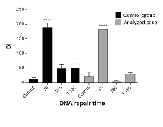
The effectiveness of the repair mechanisms did not differ between this patient and the control group (Figure 1). Immediately after irradiation (T0), moderate DNA damage was observed in both, patient and control group. Additionally, damage was significantly greater that observed in controls without irradiation, and at T60 and T120 min post-irradiation (p<0.0001). Regarding polymorphism, SNPs were observed for NOS3 G894T, GSTA1 C69T, XRCC1 R399Q and TP53 R72P; the genotypes determined were G/T, C/T, A/A, C/G, respectively. No changes were observed for genes GSTP1 A313G (A/A) and SOD2 T47C (T/T). We also found a significant association between NOS3 G894T polymorphisms and grade ≥2 acute skin radiotoxicity in patients with neo-adjuvant chemotherapy in our control database, just as in the study case (OR=14.7; p=0.03).

Figure 1 Repair kinetics of in vitro irradiated lymphocytes in the patient and control groups. Damage index (DI, expressed in arbitrary units) vs the time elapsed after irradiation is shown. ****p<0.0001 vs Control, T60 (1h) and T120 (2h); one way ANOVA and Tukey’s multiple comparisons test a posteriori. Results are expressed as mean±SEM.
Clinical radiosensitivity is a dynamic process that involves the gradual evolution of complex lesions.5 Around the turn of the century, there was growing consensus that this phenotype should be considered as a polygenic feature that depended on the combined influence of several risk alleles.6 Nevertheless, this phenotype is better understood as a multifactorial trait, where other external factors participate along with genetic polymorphisms.1,6,7 In relation to these elements, we validate the association of radiotoxicity with breast size, BMI and antineoplastic treatment, in coincidence with Back et al.8 In cellular terms, unlike other authors,9 we did not detect abnormalities in the initial damage and DNA repair capacity after in vitro irradiation. The dynamics of DNA repair observed in the patient show a good kinetics of cell recovery, with a reduction in radio-induced genomic damage in T60 and similar residual damage with the control group. In reference to polymorphisms, although the state of TP53 and XRCC1 could confer some genomic instability, the influence of these genotypes on the acute radio-toxicity of our patient could not be verified. On the other hand, it is known that the activity of antioxidant enzymes can influence the effectiveness of radiotherapy and thus it is plausible that their polymorphic variants affect the development of the side effects.10 We could not corroborate this statement in relation to GSTP1, SOD2 and GSTA1 polymorphism. However, results of NOS3 G894T polymorphism agreed with previous results10 regarding the occurrence of acute radiation skin toxicity in patients with neo-adjuvant chemotherapy. In this sense, some authors mentioned that chemotherapy increased skin reactions, and treatments with anthracycline interfered in their intensity.2,10
As a corollary of our analysis and considering that radiosensitivity is a multifactorial trait, different elements must be considered together with the polygenic component. Although radiogenomics led to a better understanding of the pathways involved in the determination of this phenotype, the involvement of the tissue microenvironment forces us to evaluate this response at a broader level of complexity not yet clarified. We believe that variables such as breast volume and BMI should be considered at the beginning of treatment and subsequently integrated into a broader analysis model that includes intrinsic variables such as the individual's radiogenomic profile. The therapeutic approach is not only important for the local control of the disease but also for the implications for the patient's quality of life. It is essential to identify solid factors that predict the course of treatment in relation to its adverse effects, especially in patients at high risk of suffering unwanted effects on the skin.
To Marcelo Martínez for helping with the irradiations and Adriana Di Maggio for manuscript correction.
None.
Approved by the Committee of the Asociación de Genética Humana (AGHU), Mar del Plata, Argentina.
The author(s) confirm that this article content has no conflicts of interest.

©2019 Ocolotobiche, et al. This is an open access article distributed under the terms of the, which permits unrestricted use, distribution, and build upon your work non-commercially.