International Journal of
eISSN: 2574-8084


Case Report Volume 1 Issue 1
1Department of Radiology, Teerthanker Mahaveer University, India
2Department of Obstetrics & Gynecology, Teerthanker Mahaveer University, India
Correspondence: Arjit Agarwal, Department of Radiology, Teerthanker Mahaveer Medical College & Research Centre, Teerthanker Mahaveer University, A-46, Gandhi Nagar, Moradabad-244001 (Uttar Pradesh), India, Tel -9012209103
Received: October 30, 2016 | Published: November 10, 2016
Citation: Agarwal A, Agarwal S, Chandak S. Pilimiction in ovarian cystic teratoma: a rare and diagnostic complication of a common entity. Int J Radiol Radiat Ther. 2016;1(1):22-24 DOI: 10.15406/ijrrt.2016.01.00005
Pilimiction (passage of hair in urine) is a rare urinary complaint which a clinician comes across during his practice. The condition may appear absurd in first impression but has a great clinical and diagnostic significance. Mature Ovarian teratoma/ovarian dermoids are one of the most common pelvic masses, which commonly manifests with pelvic pain and abdominal distention. The most common complication is torsion of the ovary with rare events of rupture, ovarian infarction and malignant transformation. Spontaneous rupture of the ovarian dermoid into the urinary bladder is the most unexpected and least common complication. We present a case of a young female presenting with complaints of pilimiction and lower abdomen pain, where ultrasound suspected a Urinary bladder mass. The diagnosis of ovarian dermoid invading the urinary bladder with formation of ovarian cysto-vesical fistula was made on the basis of CT and MRI. The event simulates the overgrowth of the placenta percreta into the urinary bladder; hence can be depicted as ovarian cystic teratoma percreta.
Keywords: dermoid, pilimiction, hair, fistula, ovary, teratoma
Ovarian dermoid is one of the most common pelvic mass in females with diagnostic imaging features. Torsion of the dermoid is known complication; however rupture with adhesion and fistulation in to the adjacent pelvic viscera is a rare phenomenon; which is described in this case. The presenting feature of passage of hair in urine is a diagnostic symptom in the described entity.
A young parous female of 30 years of age presented to the Department of Gynecology with complaint of lower abdomen pain with recent two episodes of Pilimiction (Figure 1) and burning sensation. There was no history of hematuria or colour change in urine. Menstrual cycles were regular with non complicated previous obstetric history. Clinical examination showed no major upper abdominal finding. A globular lump was palpable in the midline in lower abdomen which was non tender and with restricted mobility. Per speculum and per rectal examinations were unremarkable.
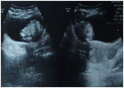
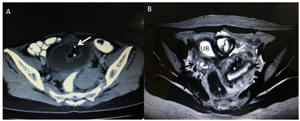
There was a pelvic ultrasound done by a local sonologist which reported a mass in the urinary bladder with normally visualized both ovaries & uterus. The scan was repeated and revealed an intra vesicular echogenic mass (Figure 2) with normal right ovary and uterus. However; left ovary was not visualized. Due to non visualization of the Ovary, the patient was advised to get a pelvic MRI, which revealed a mixed intensity lobulated well defined mass along the dome of the Urinary bladder with both intra & supra vesicular components (Figure 3). There were internal T1 hyper intense areas showing low intensity on fat suppression and a focus of signal void; suggesting internal fat and calcific intensities, respectively. Few small cyst like foci (follicles) could be visualized in the supra vesicular component of the mass with non visualized separate left ovary. Right ovary and Uterus were normal. Diagnosis of Left Ovarian dermoid cyst with cysto-vesical fistula was made. In view of academic interest and for diagnostic correlation, pelvic CT scan was acquired; under complete patient satisfaction and informed consent (Figure 4).

Figure 2 Pelvic trans abdominal ultrasound image showing echogenic mass in the urinary bladder lumen with normal uterus.

Figure 3 Pelvic MRI showing a well defined supra and intra vesical mass with breech in the dome of the bladder. (A) Axial T1 W image & (C) Sagittal T1 W image showing intra luminal bright area (shown by white arrow) with corresponding suppression on STIR (B & D) images s/o fat intensity.

Figure 4 (A) Pelvic CT scan axial image showing intra vesical lesion of fat attenuation (asterisk) and focal calcification.(white arrow).
(B) T2 W Axial MRI pelvis image at the level of dome of the Urinary bladder (UB) is showing left ovarian tissue contained dermoid and peripheral normal follicles (white arrows). Right ovary (black arrow) and Uterus (astersik) are normally visualized.
Pre-operative cystoscopy was done, which revealed intra luminal mass projecting from the dome region with dense urine and floating hair/sebaceous material. The patient underwent laparotomy and a left ovarian dermoid of size 8.5x6.5cms was seen reaching anterior & superior to the Urinary bladder with torsion of the infundibulopelvic ligament. The cyst was showing surrounding inflammation with invasion into anterior superior wall of the Urinary bladder. Left sided salpingo-oophorectomy was done with partial cystectomy and bladder repair. There was trans mural infiltration of the bladder layers i.e. serosa, muscularis and mucosa. Histopathology of the specimen revealed non-specific chronic inflammation of the bladder mucosa with features of mature cystic dermoid of the left ovary. Post operative status was normal with resolution of the urinary complaints.
Dermoid cyst is a mature benign form of teratoma, with bimodal age of the presentation; accounting for approximately 20% of all ovarian neoplasms.1 Most of the dermoids are asymptomatic and discovered incidentally on pelvic examination or ultrasound. 15-20% of the cases present with ovarian torsion.2 Other uncommon complications include rupture, malignant transformation (1-2%), infection and rarely invasion into adjacent viscera and autoimmune hemolytic anemia (<1%).3 Spontaneous rupture and infiltration into the adjacent viscera is a rare event simulating percreta type of the invasive placental tissue with Urinary bladder being the most common.
The dermoid consists of tissue derived from all the three germ layers forming a dermoid mesh of sebaceous material like hair tuft, tooth, bones and neural elements. Hence Pilimiction is a diagnostic clinical symptom indicating the ruptured ovarian dermoid with cysto-vescial fistula formation. Patient may complaint of hematuria, pyuria and strangury.4
Literature search highlights the sequence of events behind the fistulous communication in between dermoid cyst and visceral structures based on leak-adhesion-fistulation theory. The laparatomy finding shows ovarian torsion as the primary event leading to focal ischemic necrosis of the dermoid causing release of the sebaceous material, chemical irritation and its adhesion with adjacent organ, terminating into a fistulous communication.5 A case of ovarian cysto-enteric fistula is reported with adhesions as the primary event.6
Ultrasound shows an echogenic mass in the ovary, however in cases of infiltration into the bladder, primary bladder tumour and large vesical calculus creates diagnostic dilemma.7 Computed tomography is the imaging modality of choice because of unquestionable demonstration of the calcification and fat within the mass. However; the ovarian tissue cannot be visualized with confirmation. In the case presented, MRI is of great diagnostic significance due to the visualization of the normal ovarian follicles in the vicinity of the dermoid cyst, obviating its ovarian origin. T1 hyper intense areas of fat and voids of calcification are convincingly appreciated on routine MRI sequences.
Salpingo-oophorectomy with partial/conservative bladder resection and repair is the primary surgical procedure which is curative. Histopathological diagnosis is required to rule out any signs of malignant transformation.
Pilimiction is a clinical symptom which is best correlating with the radiological features of dermoid cyst and a diagnostic pointer for the clinician and the radiologist. Imaging is useful in operative planning with focus on site of origin of the dermoid, size of the cyst, communication with viscera and possibility of torsion.
None.
Author declares that there is no conflict of interest.

©2016 Agarwal, et al. This is an open access article distributed under the terms of the, which permits unrestricted use, distribution, and build upon your work non-commercially.