International Journal of
eISSN: 2573-2889


Review Article Volume 3 Issue 3
Citizen Scientist, USA
Correspondence: Abraham A Embi, Formerly Affiliated with VA Hospital, University of Miami, 13442 SW 102 Lane, Miami, Florida, USA, Tel +305 505 4979
Received: May 06, 2018 | Published: May 24, 2018
Citation: Abraham AEBS. The secondary role of UV light in swimmers melanoma genesis. Int J Mol Biol Open Access. 2018;3(3):121-123. DOI: 10.15406/ijmboa.2018.03.00064
Background: Swimmers are known to have a higher incidence of cutaneous melanoma. Experimental studies are unable to explain the role of ultraviolet radiation (UV) as the sole factor in swimmers cutaneous melanomas. Other mechanisms, such as the prevalence of the decomposition of Hydrogen Peroxide (H2O2) in the hair follicles has been hypothesized by several authors as another etiological factor. For example, the H2O2 generated by keratinocytes diffuses into melanocytes and may play an important role in the etiology and pathogenesis of cutaneous melanoma. Other in vitro experiments document that in water submerged hairs, sites such as the infundibulum and at wounded hair follicles is where exogenous H2O2 could penetrate. The energy released during the decomposition of H2O2 by catalase in the eukaryotic cell was also hypothesized to be an additional factor for the origin of cancer. Epidemiological studies attribute the low incidence of Melanoma genesis in countries such as Japan to the beneficial effect of the consumption of the seaweed, Sargasum filipendula, which has antiproliferative and antioxidant properties. This manuscript supports H2O2 as being the missing attributable risk in swimmers cutaneous melanomas.
Methods: A literature survey was performed, which aimed to identify exogenous H2O2 as a key factor in the causation in cutaneous swimmers Melanoma genesis.
Results: In Swimmers, all published theories as well as actual in vitro experiments identify H2O2 as essential factors for cutaneous melanoma. In water-submerged hairs, there are two sites where exogenous H2O2 can penetrate the human hair follicle: At the hair shaft/skin junction, and sites where injury has damaged the external follicular wall.
Conclusion: Published evidence demonstrates that the incidence of cutaneous melanomas is higher in swimmers that in the general population. H2O2 forms by the conversion of dissolved organic matter by the sun UV light in fresh or salt-water bodies. In Swimmers, spontaneously penetrating into the hair follicles. Melanoma tumor cells are then formed by the decomposition of H2O2 by catalase; and the malignant cells then spread into the surrounding tissues. The sun UV light playing a secondary role with H2O2 hypothesized as the primary attributable risk.
Keywords: biophysics, cancerogenesis, melanoma hair follicle, H2O2 decomposition, Cancer UV
Melanoma is known since the ancient times and has long troubled caregivers, primarily due to its heterogeneity.1 Swimming in fresh or salt-water are linked to increase risk for cutaneous melanomas.2 Experimental studies are unable to explain the role of ultraviolet (UV) radiation as the sole factor in Melanoma genesis. It has been hypothesized that one of the factors for melanoma tumors could be the generation of reactive oxygen species [namely Hydrogen Peroxide (H2O2)] by melanin “may play an important role in the etiology and pathogenesis of cutaneous melanoma”.3,4
H2O2 from keratinocytes penetrates melanocytes
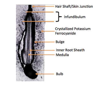
Published reports identify keratinocytes as the main source of H2O2 penetrating melanocytes. “Basal hydrogen peroxide (H2O2) levels in normal human epidermal keratinocytes (NHEK) and melanocytes (mel) were compared on a per cell basis and found to be significantly higher in keratinocyte. Because the ratio of keratinocytes to mel in skin is 36:1, keratinocytes may act as a source of reactive oxygen species (ROS) even by passive diffusion and, thus, affect melanocytic functions”.5 Melanin has been described as a photo protective factor in the skin;6 it could also act as double edge sword. How? Melanin besides functioning as a broadband UV absorbent is a reservoir of toxic H2O. In addition, experiments have documented the infundibulum (Figure 1) (Figure 2) & Video recording and the wounded follicles (Figure 3) & Video recording) as sites where exogenous H2O2 penetrates the water immersed hair follicles; and is the region where melanoma tumor cells have been previously reported to form.7,8
Additional experiments supporting H2O2 as “missing attributably risk”
Besides the above-mentioned molecular factors, a biophysical hypothesis was proposed linking energy derived from the decomposition of H2O2 by catalase in eukaryotic cells to be an additional factor for the origin of cancer.9
Worldwide epidemiological reports, antioxidant diet and melanoma
Circumstantial evidence attributes the low incidence of Melanoma genesis in Japan to the beneficial effect of the consumption of the brown seaweed, Sargasum filipendula. Polysaccharides from Sargasum filipendula were found to have antiproliferative and antioxidant properties.10,11
A literature survey was conducted, which demonstrated that the carcinogenic H2O2 molecules penetrate the hair follicle at the junction of the skin and hair shaft at the infundibulum, and through the injured external wall.
Sites of melanoma genesis in hair follicles
Published literature supports the fact that hair follicles are anatomical sites where the melanoma tumor cells are formed. As shown in Figure 1 & Figure 2, the spontaneous penetration of exogenous H2O2 molecules only occurs through the infundibulum of the hair follicles. Injuries to the outer wall of hair follicles induce the penetration of H2O2 molecules into the follicle (Figure 3). This observation was consistent and was confirmed by a study, which reported “Water H2O2 Levels as factor in swimmers melanoma”.12

The decomposition of H2O2 as key factor in melanoma genesis
A novel paradigm was hypothesized by recent experiments that demonstrated the elevation of ROS due to melanin, which served as a redox generator, for which the authors stated that, “this may play an important role in the etiology and pathogenesis of cutaneous melanoma”. In another recent experiment that mimicked the Swimmers’ water environment, H2O2 was also identified as penetrating the infundibulum of the hair follicles. In both cases, H2O2 was identified as a possible cause for the formation of melanoma tumors in the hair follicle. The evidences justify the role of H2O2 molecules as the elusive attributable risk in swimmers melanoma3XXXX, UV radiation playing a secondary role, since its primary role only concerns the conversion of the dissolved organic matter present in water into H2O2 molecules, as shown in the panel below (Figure 4).13

Etiology of swimmers melanoma genesis
Based on the evidences found in recent experiments and published literature, it is hypothesized that melanoma tumor cells originate and spread primarily from the hair follicles, resulting from the decomposition of H2O2 by catalase. The role of UV light being secondary.
Further Research is warranted
This author recommends for experimentations on skin for the animals of choice to be pig or mice, since their skin and hair follicles mimic those of humans.14 In pigs it was also observed “Melanocytic cutaneous proliferation appeared to begin the hair follicle and spread to adjacent tissue”.15 Studies in mice revealed that “Wounding mobilizes hair follicle stem cells to form tumors”.4 Experiments performed in pigs demonstrate that “Melanocytic cutaneous proliferation (melanomas) appeared to begin in the hair follicle and then spread into surrounding connective tissue, sweat glands, sub dermal connective tissue and fat”, which raises a few questions. The first question that arises is: Would the topical application of antioxidants in pig inhibit the generation of melanoma or affect tumor regression? This question arises since pigs cool their temperature and protect their skin by bathing in mud. Dermatological studies supports and justifies the topical application of antioxidants to reduce dermatological lesions. The topical application of antioxidants is rationalized so as to avoid generating systemic imbalances of ROS and enzymatic systems. The study reports, “In situations where this balance is broken, various cellular structures, such as the cell membrane, nuclear or mitochondrial DNA may suffer structural modifications, triggering or worsening skin diseases”.16 The second question that arises is, in the paper published in PNAS by Wong Could the prevalence of H2O2 molecules at the site of injury be ruled out in Melanomagenesis?17A negative answer is supported by experiments where “Once a skin wound occurs, based on an experiment performed on zebra fish by mechanically injuring its tail fin, a sustained rise in H2O2 concentration was detected at the wound margin immediately after the injury occurred”.4 For the sake of a hypothetical inquiry, supposing that the in vivo experiments in pig and mice were executed and would turn out to be successful, the results would serve as a foundation for validating H2O2 as the elusive attributable risk in Melanoma genesis. The role of UV light would remain as secondary.
None.
None.

©2018 Abraham. This is an open access article distributed under the terms of the, which permits unrestricted use, distribution, and build upon your work non-commercially.