International Journal of
eISSN: 2573-2889


Mini Review Volume 2 Issue 3
1Department of Physiology, Faculty of Pharmacy, University of Granada, Spain
2Department of Physical and Analytical Chemistry, University of Oviedo, Spain
Correspondence: Carlos López-Chaves, Department of Physiology, Campus Cartuja, University of Granada, 18071 Granada, Spain, Tel 0034 665701175
Received: April 19, 2017 | Published: May 9, 2017
Citation: López-Chaves C, Alvaredo JS, Bayón MM, et al. Gold nanostructures in biomedical technology. Int J Mol Biol Open Access. 2017;2(3):105-106. DOI: 10.15406/ijmboa.2017.02.00024
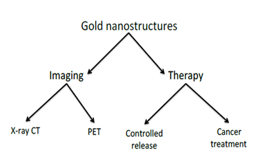
Nanomaterials have been defined as any material possessing at least one dimension in the nanometer scale between 1 and 100nm. As a result, a new field has emerged during last decades: Nanotechnology. It can be defined as the design, synthesis and application of materials and devices whose size and shape have been engineered at the nanoscale. In addition to the main properties of Nanomaterials, Au nanostructures exhibit a variety of optical, physical and chemical properties (localized surface plasmon resonance, photoluminescence, surface-enhanced Raman scattering, resistance, bioinert nature or photo thermal effect) that make them especially interesting for biomedical applications. Here, we will summarize those with high relevance and impact on the future of the medicine in terms of development of new detection and imaging techniques and innovative therapies against cancer.
Keywords: gold, nanomaterials, nanotechnology, nanomedicine
Gold is mainly known in the jewellery industry due to its tone, rarity and value. However, there are many others advantages (such as bioinert properties1 or photo thermal capabilities)2,3 that make gold an important material for biomedical applications. Indeed, new therapeutic methods are now being developed by combining nanotechnology and gold.4

X-ray computed tomography (X-ray CT)
This technique is one of the most widely used modalities for medical imaging. It provides anatomical information for diagnostics in a cheaper way than other methods. However, the contrast between different types of soft tissues is currently impossible for conventional X-ray CT, so that a good contrast can only be achieved between hard and soft tissues.5,6 As a result, conventional X-ray CT cannot be used alone to differentiate cancerous from normal tissues, rendering it essentially useless for the early detection of cancer or cancer metastasis.
As a response to this inconvenient, and given that the attenuation of X-rays by tissue depends on the electron densities, Au provides a strong contrast relative to most of the naturally occurring elements found in the body.7 The presence of Au nanostructures can greatly enhance X-ray attenuation, creating high contrast on the CT image. Furthermore, gold nanostructures have shown prolonged presence in the circulation, lower renal toxicity, and well-established surface chemistry for the conjugation of targeting ligands. All these attributes make Au nanostructures promising contrast agents for clinical X-ray CT applications.8,9
Positron emission tomography (PET)
PET is one of the most frequently used modalities in molecular imaging, and can be employed to collect information about the expression of molecular species on a biological tissue. It is based on the detection of pairs of gamma rays emitted indirectly by a positron emitting radionuclide, which is introduced into the body on a biologically active molecule. Thus, by chelating or covalently attaching the radionuclide to the surface of Au nanostructures, doctors are able to visualize those areas where gold nanostructures are accumulated.5,10-12
Besides, it is used to obtain valuable information about cellular function and gives insight into the molecular processes of a living organism, enabling diagnosis even before changes at any anatomical are observed.
In addition to this, both techniques may also be combined for multimodality imaging; which gives the opportunity to obtain better results regarding biodistribution, targeted accumulation and clearance of Au Nanomaterials by joining the morphological and quantitative data acquired from X-ray CT and PET.13-15
Drug loading and controlled release
Being able to load and release a drug in the target tissue (thus considerably reducing side effects), supposes a major step in the development of a medicine. To this purpose, several methods have been published: Recent studies have been focused on the development of so called Au hollow nanostructures. In this way, drugs can be loaded in the interior of these nanostructures to decrease the degradation and increase loading efficacy. Furthermore, by modifying the surface, authors have been able to deliver the drug under controlled circumstances (pH, temperature-sensitive polymer) or tissues (by attaching specific antibodies to the surface).16
Additionally, drugs can also be loaded onto the surface of the gold Nanomaterials. For instance, the high affinity between Au and other atoms like sulphur or nitrogen, the electrostatic interactions or van der Waals forces as well as hydrogen bonding, have showed promising results as tools for drug delivery.17,18 In any case, gold nanostructures have shown promising properties in realizing drugs, based on their ability to heat and break chemical bonds between the drug molecule and the Au surface under the irradiation of light.1920 Thus, by combining these techniques, researchers have been able to develop a smart gold nanostructures able to load the drug, travel to the target tissue and deliver the drug.
Cancer therapy
Based on the ability of gold nanoparticles to heat under light irradiation, photo thermal therapy has gained importance last year’s.2,21 This process relies on the use of hyperthermia to kill tumour cells (after exposing them to high temperatures for several minutes). However, the nonspecific heating which also destroys adjacent tissues implies a counter indication in this therapy. The fact that these hyper thermal agents are developed in the nanoscale significantly reduces the side effects. Furthermore, it is possible to accumulate gold Nanomaterials at the tumour zone thanks to active targeting enabled by ligands such as antibodies.22,23 Thus, the accumulation of Au nanostructures at the desired site will lead to selective heating of target cells after laser irradiation, minimizing potential damage to the surrounding tissue. This advantage, together with the fact that Au nanostructures can serve as contrast agents for various imaging techniques, would allow physicians to achieve an imaging-guided therapy.
Being able to quickly detect and treat a disease like cancer with minimal counter indications is a goal that scientists have been pursuing for several years. The huge potential benefit of Au Nanomaterials in the development of nanotechnology and nanomedicine, has contributed to the increased use of these structures as feasible tools in new imaging techniques, drug release and cancer treatment.
The authors gratefully acknowledge the financial support from the Spanish MICINN (Spanish Ministry for Science and Innovation, Grant Number CTQ2011-23038) and MECD (Spanish Ministry for Education, Culture and Sports, Grant Number FPU13/00062). The authors thank JH Thompson for translating the manuscript into English.
Author declares that there is no conflict of interest.

©2017 López-Chaves, et al. This is an open access article distributed under the terms of the, which permits unrestricted use, distribution, and build upon your work non-commercially.