International Journal of
eISSN: 2573-2889


Review Article Volume 2 Issue 2
Donetsk Physical and Technical Institute National Academy of Science of Ukraine, Ukraine
Correspondence: Donetsk Physical and Technical Institute National Academy of Science of Ukraine, 03680 Kiev, Nauky av. 46, Ukraine
Received: February 19, 2017 | Published: April 12, 2017
Citation: Grebneva HA. A polymerase tautomeric model for radiation induced bystander effects: a model for untargeted base substitution mutagenesis during error prone and SOS replication of double stranded DNA containing thymine and adenine in rare tautomeric forms. Int J Mol Biol Open Access. 2017;2(2):63-74. DOI: 10.15406/ijmboa.2017.02.00018
Currently, untargeted mutations are studied in the context of bystander effects. Up to now the nature and mechanisms of formation of untargeted mutations is poorly understood. Untargeted base substitution mutations are base substitution mutations then one or some nucleotides are inserted in DNA molecule on, so called, undamaged sites of DNA. Here the polymerase-tautomeric models of ultraviolet mutagenesis and bystander effects are developed. The mechanisms of untargeted base substitution mutations formation caused by cis-syn cyclobutane thymine dimers and bases pairs of adenine-thymine or thymine-adenine in rare tautomeric forms that are in small neighbor from any cyclobutane dimers are proposed. Five rare tautomeric forms may form for thymine and adenine. These rare tautomeric forms will be stable if corresponding nucleotides are part of cyclobutane pyrimidine dimers or are in small neighbor of the cyclobutane dimers and during DNA synthesis. Structural analysis of bases incorporation shown that adenine in rare tautomeric form A1* may result in untargeted homologous transversion Т-А→А-Т. Thymine in rare tautomeric form T1* may result in untargeted transition А-Т→G-C or untargeted homologous transversion А-Т→Т-А. Molecules of the thymine in rare tautomeric form T4* may result in transversion A-Т→С-G only. Thymine in rare tautomeric form T5* may result in untargeted transversion А-Т→С-G or untargeted homologous transversion А-Т→Т-А. Thus, molecules of adenine and thymine in definite tautomeric forms are in small neighbor from any cyclobutane pyrimidine dimers may result in untargeted base substitution mutations.
The major underlying cause of mutations in cancer is DNA damage.1 The formation of mutations is the main cause of genetic diseases and cancer, as well as the major cause of aging.2 Understanding of processes that occur during the formation of mutations, it is necessary for the developing of strategies and methods to combat cancer, as well as for the development of prevention methods that would contribute to a decrease in the probability of occurrence of cancer and hereditary diseases.
Radiation-induced genomic instability is a modification of the cell genome found in the progeny of irradiated somatic and germ cells but that is not confined on the initial radiation-induced damage and may occur de novo many generations after irradiation.3‒5 It has been shown that radiation can, by itself, induce a type of genomic instability in cells, which enhances the rate at which mutations and other genetic changes arise in the descendants of the irradiated cell after many generations of replication.4 Genomic instability leads to the formation of malignant tumors. Genomic instability is the process whereby gene mutations increase. Although radiation-induced genomic instability has been studied for years, many questions remain to be answered.6 Delayed mutations and untargeted mutations are two features of genomic instability.7 In recent decades untargeted and delays mutations are combined in bystander effects.8‒10 Genomic instability and the bystander effect have been linked experimentally.11 It is believed now that untargeted and part of delayed mutations appears on not damaged DNA sites.7 There are now known to be many late expressed effects of exposure that cannot simply be explained on the basis of direct ionizing radiation DNA damages. Examples include genomic instability, bystander effects and adaptive responses.12 The bystander effects are defined as the induction of cellular damage in un irradiated cells, induced by irradiated cells in the surrounding area.8
Six mechanistic explanations for the phenomenon have been proposed. The most convincing explanation of radiation-induced genomic instability attributes it to an irreversible regulatory change in the dynamic interaction network of the cellular gene products, as a response to non-specific molecular damage3 Campa et al. underline the central role of cell-cell communication on non-targeted effects.13 According Averbeck the discovery of non-targeted and delayed radiation effects has challenged the classical paradigm of radiobiology.14 Watanabe assume that a radiation cancer-causing target is protein.15 Wright assume that radiation-induced genomic instability in vivo may not necessarily identify gnomically unstable somatic cells but the manifestation of responses to ongoing production of damaging signals generated by genotype-dependent mechanisms having properties in common with inflammatory processes.16 Furthermore, it is still not known what the initial target and early interactions in cells are that give rise to non-targeted responses in neighboring or descendant cells.10 Morgan believes that it is unlikely that these untargeted effects are directly induced by cellular irradiation. Instead, it is proposed that some as yet to be identified secreted factor can be produced by irradiated cells that can stimulate effects in no irradiated cells (death-inducing and bystander effects, clastogenic factors) and perpetuate genomic instability in the clonally expanded progeny of an irradiated cell. Furthermore, it must have the potential to stimulate cellular cytokines and/or reactive oxygen species17 Lyng et al.11 suggest that initiating events in the apoptotic cascade were induced in unit cells by a signal produced by irradiated cells and that this signal can still be produced in the progeny of irradiated cell.11
Moreover, it is concluded that only a third of the variation in cancer risk among tissues is attributable to environmental factors or inherited predispositions. The majority is due to “bad luck,” that is, random mutations arising during DNA replication in normal, noncancerous stem cells.18 The conventional paradigm relates the reason of mutations exclusively to sporadic errors of DNA polymerases.19‒23 (a more detailed review, see in ref.)24 The reasons of mutation origination are explained by the so-called “A-rule”.22,25 It is assumed that DNA polymerases insert non-complementary bases opposite the damages.19,22 As part of the polymerase model mechanisms of the targeted,26,27 and untargeted19,20,28‒33 base substitution mutations have been proposed. The mutations that result from incorrect bases are often targeted; that is, they occur at the same position as the photoproduct.26,27 Sometimes mutations are formed in the vicinity of damage, a process that is termed untargeted mutagenesis19, 20, 28-33.Untargeted mutations currently called mutations appearing on so-called "undamaged" DNA sites.19,20,28‒36 Untargeted mutagenesis requires the same proteins that are required for translesion synthesis.34 Untargeted mutagenesis occurs at SOS replication and error-prone DNA synthesis.19 Untargeted mutagenesis is characterized by a high rate of education of transversions.19,34,36 It is assumed that untargeted base substitution mutations formed on undamaged DNA sites in result in random errors of DNA polymerases.19,20,28‒32,37‒39
Many specialized DNA polymerases can lead translesion synthesis opposite of cyclobutane pyrimidine dimers 23,31,35,40‒44 and participate in the formation of untargeted mutations. For example, the DNA polymerase pol θ is processivity polymerase, which implies that it can strongly regulated to avoid mutagenesis.41 Error-prone polymerases V Escherichia coli (Pol V) and polymerase IV (Pol IV) are responsible for SOS-induced untargeted mutations.23,35,42 DNA polymerase IV causes untargeted and the targeted SOS mutagenesis.31,43 It can produce of the mutation in the vicinity of the damage those results in untargeted mutagenesis.19
In recent decades, the main efforts were focused on experimental determination and computer modeling of the structure of DNA polymerases and the mechanisms of nucleotide incorporation in the active sites of polymerases.45‒56 Structural data are obtained for the mechanisms of the targeted and untargeted mutagenesis formation under the translesion synthesis via cyclobutane pyrimidine dimers.57‒59 The role of the polymerases in cancer is investigated.59,60 However, despite the obvious advances in the study of the structure and functions of polymerases 19,20,37,39‒41,45‒55 the modern theory of mutagenesis, based on polymerase paradigm, cannot fully explain many phenomena of mutagenesis. These include the reasons for the formation of hot and cold spots of ultraviolet mutagenesis.1,62 Much of it is not clear in the nature and mechanisms of formation of delayed mutations.63‒66 It is not entirely clear mechanism for the formation of complex mutations.67,68 It is not clear why some cyclobutane pyrimidine dimers do not cause mutations, while others, seemingly the exact cause of transition or transversion mutations, or the just the same cyclobutane pyrimidine dimers cause frame shift mutations. The mechanism of formation the targeted frame shift mutations does not clear.69‒73 Even the mechanisms of formation of the targeted base substitution mutations are not entirely clear. It is not clear why in some cases there are transitions, while in others cases there are, for example, homologous transversions, although it would seem that cause their cis-syn cyclobutane pyrimidine dimers exactly the same. It is not entirely clear how the mutations occur in the repair, such as excision repair or postreplicative repair.74,75
There remains much uncertainty on the issue of the nature and mechanisms of formation of untargeted mutations. It is not entirely clear why mutations occur on some "not damaged" sites of DNA. What's the difference between these sites from those on that do not appear untargeted mutation? As part of the polymerase model answer is no different. But is it? It is not clear why sometimes there are untargeted base substitution mutations, and, in other cases, untargeted frame shift mutation frame shift. When there are untargeted base substitution mutations it is not clear why in some cases appear untargeted transitions, in other cases appear untargeted transversions. There is no model explaining the mechanisms of untargeted complex mutations. And, of course, it is not as clear as may appear untargeted delayed mutations. It is known that untargeted and delayed mutations are a major manifestation of genomic instability.76 Instability of genome is the main cause of cancer.77 It is not clear why, in the genomic instability that leads to cancer, increases dramatically the number of untargeted and delayed mutations. In other words, the evidence suggests that the nature and mechanisms formation of untargeted mutations, genomic instability and cancer cannot be completely understood. Therefore, it is important to examine the nature and mechanisms of untargeted mutations in more detail.
I have attempted to construct a polymerase-tautomeric model for UV-induced mutagenesis,24,78‒99 based on idea by Watson and Crick77 that changes in tautomeric state are possible for DNA bases. It is proposed a mechanism for changes in the tautomeric state of base pairs under ultraviolet irradiation of DNA.79‒82 It was assumed that the tautomeric state of the constituent bases may change during the formation of cyclobutane pyrimidine dimers.80‒82 Five new rare tautomeric conformations of the adenine and thymine,81,82 they shown on Figure 1, and seven of the cytosine and guanine.80,83 Such rare tautomeric forms of DNA bases are stable when they are parts of the Cis-syn cyclobutane pyrimidine dimers and they are stable under DNA synthesis.81‒84
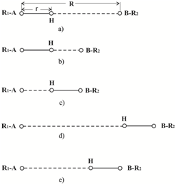
Figure 1 Diagrammatic representation of hydrogen bond between molecules R1-A and B-R2:
a) r-length of a valence bond between A and atom of hydrogen H. R - length of hydrogen bond. b) the molecules bound by the hydrogen bond were pulled together so, that the atom of hydrogen is almost at the center between the molecules c) the atom Н has formed the same strong bond with BR2 as with R1A d) hydrogen bond has got longer. Н is connected by a valence bond with BR2 e) length of Н-bond was normalized, the atom of hydrogen now is connected. by a strong valence bond, to a molecule BR2.
It is proposed a polymerase-tautomeric model for the mechanisms of targeted base substitution mutations formation during error-prone or SOS synthesis of DNA containing Cis-syn cyclobutane pyrimidine dimers 24,84‒87,98 It is developed a polymerase-tautomeric model for targeted frame shift mutations, insertions98,89,98 and deletions90,91,98 caused by Cis-syn cyclobutane thymine dimers. It is proposed a polymerase-tautomeric model for the mechanisms of targeted complex insertions92,98 and a polymerase-tautomeric model for the mechanisms of hot and cold spots of ultraviolet mutagenesis.93 It is proposed a polymerase-tautomeric model for the mechanisms of targeted delayed base substitution mutations caused by Cis-syn cyclobutane thymine dimers.97 Polymerase-tautomeric model for untargeted substitution mutations formation when DNA molecule contains Cis-syn cyclobutane thymine dimers also have been developed.94‒96 However, preliminary works on untargeted mutagenesis94,95 relied on a number of hypotheses, from which in the future, under the study of the nature of the targeted mutagenesis I refused.84
In order to understand how untargeted mutations are formed, it is necessary to understand how DNA damages are formed, which can be a source of untargeted mutations. For this, first, it is necessary to examine the processes by absorption of excitation energy in the DNA molecule. Secondly, we must look to which the chemical changes of DNA structure it will lead. Thirdly, it is necessary to understand the conditions under which the damages of DNA will be stable. Fourthly, to explore how these changes in the DNA can result in mutations under the replication and repair. In the first place I'll look at how the tautomeric state of bases DNA may change under UV light.
It is developed a model of the mechanism changes of the tautomeric states of the base pairs of DNA. It has been proved in detail for the case when the DNA molecule is irradiated with ultraviolet light.79‒83,98,99 After irradiation of DNA by ultraviolet light excitation energy ultimately localized on one of the bases. This results in the excitation of electronic-vibration states.100 For a triplet level excitation, the most probable process for relaxation involves the transformation of energy to oscillations of the neighboring atoms,10 in other words in a heat. No radiative deexcitation in DNA molecules occurs in a small volume of 3-5 bases pairs.102,103 This results in a strong "local heating-up", followed by initiation of normal oscillations of the bases. After several oscillations, taking ~10-14 - 10-12 sec,104‒108 the vibratory system will reach equilibrium. The oscillations of atoms will cause changes in the distances between the paired bases, in other words, in the lengths of H-bonds. Hydrogen bond is called such a bond when there is a hydrogen atom between the two electronegative atoms. The shapes of the potential curves for the protons of all three H-bonds of Watson and Crick’s G:C base pair for several lengths of H-bonds were calculated in ref.109‒108110 Calculations were based on semi-empirical potential function, capable of adequately describe hydrogen bonds with lengths different from the equilibrium lengths.111‒108 It turned out that a decrease in the length of a Н-bond by 0.02 nm from the equilibrium transforms the proton potential into a single-well. With an increase in Н-bond length, the second minimum becomes more and more obvious. The dynamics of the changes in the shape of proton potential for a wide spectrum of Н-bond lengths were calculated.110‒108 Hydrogen bonds that are formed between the DNA bases have the following property. They are characterized by a strong valence bond with one of the partner atoms in the Н-bond, and a weak bond with the other (Figure 1a). When the Н-bond length (R) of a valence bond changes, the length (r) changes very little. But distance from the hydrogen to the second atom (R-r) varies considerably.111‒108 Let hydrogen bond length (R) reduced so that hydrogen was almost in the center of the hydrogen bond (Figure 1b). In this case, it forms a strong bond with the two electronegative atoms (Figure 1c). Therefore, when the hydrogen bond begins to lengthen, hydrogen may remain in the new position (Figure 1d). After that hydrogen bond length is increased a hydrogen atom will be in a local minimum.104‒108,106‒108,101‒108 The assumption of favorable conformations is possible, as the lifetime of a triplet state is ~10-6 sec.101‒108 The lifetime of the excited Н-bond is ~4 x 10⁻⁹ sec.104‒108,113‒108 In the same time the characteristic periods of atomic oscillations are 10–14- 10–12 sec.104‒108 Therefore, there will be not more than several 10s of oscillations up to 100 oscillations that influence the length of the H-bond. This results in a time of ~ 10-10 sec for any one conformation which is much less than the lifetime of the triplet state. Thus, the hydrogen atom will remain with their partner on hydrogen bond (Figure 1e). The calculations104‒108,110‒108, 104‒108,113 semi empirical potential function for protons of hydrogen bonds was used, it was developed by Tolpygo & Grebneva.109 The proposed model is in a good agreement with the results obtained by other authors.114‒116 The transitions of the protons in hydrogen bonds are very common. Proton transitions in H-bonds occur in acids and bases, in crystals, proteins, molecular membranes, enzymes, and in other systems.117
In a canonical DNA molecule, conformational fluctuations of different DNA sites result in different metastable states.118,119 The exposure of bases from a double spiral to the solution is known as “opening” or “melting” of Watson and Crick’s pairs. In this case, the hydrogen bonds between DNA bases are broken.118 Metastable states developing with incomplete opening of the pair and with partial conservation of H-bonds and with the amine group free from H-bond are called semi open metastable states.118 There exist several models of semi open metastable states.118,119 In my opinion, the model developed by Hovorun119 is well-grounded; it explains a number of phenomena and is confirmed by experimental data. It is rather probable that the process of tautomeric state change goes in two stages. At the first stage, the metastable semi open DNA states predicted in Ref119 are formed. At the second stage, the formed rare metastable tautomeric states transform into the stable ones. Allowance the predicted119 a metastable state leading to that rare tautomeric forms of thymine T*4 and T*5 and rare tautomeric forms of adenine А*4 and А*5 (Figure 2)82,83 can be formed.
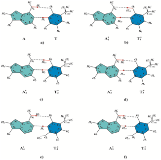
Figure 2 Possible rare tautomeric state of thymine and adenine:
a) adenine (A) and thymine (T) in canonical tautomeric forms.
b) f) molecules of adenine (Ai*) and molecules of thymine (Ti*) in rare tautomeric forms, i = 1¸5.
Double-stranded DNA contains the guanine-cytosine and adenine-thymine base pairs. In these pairs bases are interconnected by hydrogen bonds, and they are located in different strands. As a result of changes in the lengths of the hydrogen bonds caused by the strong thermal vibrations a hydrogen atom can remain with his partner in a hydrogen bond. If such an event occurs, it means that the tautomeric states of DNA bases are changed. Thus tautomeric states always change in both bases bonded by hydrogen bond because hydrogen is separated from the one base and joined to another base of the pairs.80‒82 As shown in the quantum-mechanical calculations, in all cases (except for one) hydrogen atoms are returned to the original position.120‒122 In other words, the bases are usually returned to their canonical tautomeric states.
The reasons for the stability of the rare tautomeric forms of DNA bases
Let us see under what conditions the rare tautomeric forms of the DNA bases will be saved. The rare tautomeric forms of bases are stable at Cis-syn cyclobutane pyrimidine dimers formation and in DNA synthesis.82,84 If under the formation of the Cis-syn cyclobutane pyrimidine dimer tautomeric states of its constituent bases has changed, such rare tautomeric states will be stable.82,84 The rare tautomeric forms of bases are stable because, at Cis-syn cyclobutane pyrimidine dimers formation, the DNA strand is bent and the hydrogen bonds between the bases are significantly weakened or are broken.123‒126 When the hydrogen bond becomes weaker it becomes longer. In this case, there is a second minimum and the second minimum deepens.105,106,110 Therefore, if under the thermal relaxation of the excitation energy the hydrogen atom will go to partner for the hydrogen bond, the new tautomeric state will be stable even with a small elongation of the hydrogen bond. And, of course, such a rare tautomeric state will be stable at breaks of hydrogen bonds. That is why the hydrogen atoms between the bases located in different DNA strands, which form a pair, will not be able to return to their former partners in hydrogen bonds, and will remain in the new provisions. This means that there is change of tautomeric states of bases, and it will be stable.80‒84
But the bases in rare tautomeric forms are stable and when they are near photodimers.94‒96 DNA strand containing cyclobutane dimers, especially if there are several cyclobutane dimers located close to each other so DNA strand is bent that it forms a loop, so that the hydrogen bonds between the bases located adjacent to photodimers also are torn. Hydrogen atoms cannot return to their original position, because they are no longer bound by hydrogen bonds. Therefore, if in a small neighborhood from a cyclobutane dimer the bases in rare tautomeric form are formed, such a rare tautomeric states will be stable. An important step in improving the accuracy of DNA synthesis is the stage of identification of nucleotides and nucleotide pairs. Increased ability to distinguish nucleotides polymerases achieved in particular by removing water from active sites of the enzymes.127,128 The molecule of enzyme - is usually a very large molecule and it shuts substrate molecule that falls in the active site of the enzyme, from the influence the environment. Lack of water in the active sites of polymerases preserves tautomeric states bases involved in the synthesis, when the DNA molecule is in single stranded form.
It is possible that the DNA molecule will be in single stranded form when DNA molecule is replicated. Let us estimate, what is the probability that this bases, are in rare tautomeric forms, pass into canonical tautomeric states due to contact with water molecules. Experimental evidence indicates that the lifetime of “free” guanine, i.e. the time it takes until it interacts with a water molecule, ranges from 0.1 to 10 sec, and can be as long as 1,000 sec.129 And we are talking about guanine molecules in an aqueous medium. For a guanine, the incoming DNA strand, it is much more. Different DNA polymerases synthesize nucleotides at a rate of 10/sec to 1000/sec.19‒21,23 Suppose that during the synthesis of the DNA strand at a time will be in single stranded form. The time at which it will not be protected by the enzymes is determined by the speed of the enzymes. For DNA polymerase V, having the lowest rate of synthesis it is not more than 0.1 seconds. Since the time required for bases came into contact with the water molecule and has changed they tautomeric state, more than 0.1 seconds, we see that the probability of such a process is quite small.84The results of studies on the structure of the active centers of polymerases show that the bases in rare tautomeric forms may exist in the active centers of polymerases.130‒135 More details on this issue is set out in ref98
An important question is, how often can form bases in the rare tautomeric forms. As shown experimentally in studies of hot and cold spots ultraviolet mutagenesis, cyclobutane pyrimidine dimers appear very often.61,62 Analysis of the nucleotide sequences on which cyclobutane pyrimidine dimers are appeared, which led to the formation of targeted base substitution mutations, reveals that such cyclobutane pyrimidine dimers, are often at a distance of one, two, three or four nucleotides apart61 (Figure 3). Source of damages during the formation of cyclobutane dimers and changes of tautomeric states of bases is the same. It has strong forced oscillations with non-radiative relaxation of the excitation energy from the triplet energy level.82 To change the tautomeric state of DNA bases is needed much less of energy than for the cyclobutane pyrimidine dimers formation. Consequently, rare tautomeric forms of bases may occur more frequently than cyclobutane dimers are formed. Thus, in principle, all the bases of DNA can change its tautomeric states. Of course, we must note that thymines and adenines are much less likely to change their tautomeric states than cytosines and guanines.136 The critical question is whether these rare tautomeric states are stable. And it is entirely dependent on whether cyclobutane dimers or other damages are formed around them, and whether the DNA strand is is bent opposite of this damage. If not, then all of the hydrogen atoms return to their original positions (except one rare tautomeric form of guanine and cytosine)120‒122 and rare tautomeric states of DNA bases are not formed. If next to the bases in rare tautomeric forms will be cyclobutane dimers is every reason to remain in his rare tautomeric forms. This is because during the formation of cyclobutane pyrimidine dimer DNA strand is curved, loop occurs, hydrogen bonds are broken.123‒126 Hydrogen atoms are unable to return to their former states; they are no longer associated with their partners via hydrogen bonds.82
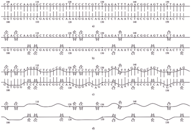
Figure 3 Formation of the loops opposite cyclobutane pyrimidine dimers at the supF site of DNA on which in Ref. 60,61 it was found hot and cold spots of ultraviolet mutagenesis:
a) Scheme of the DNA site supF which is irradiated with ultraviolet light.
b) Cyclobutane pyrimidine dimers are formed at the supF DNA site. They are marked caps.
c) Bulges (loops) are formed opposite the cyclobutane pyrimidine dimers.
Figure 3 shows a site of DNA on that is obtained hot and cold spots ultraviolet mutagenesis.61,62 The diagram shows that if the hydrogen bonds between the bases forming part of cyclobutane pyrimidine dimers are extended, the loops formed in the vicinity of these dimers. And, therefore, all rare tautomeric forms of the bases will be sustainable.
The mechanism of rare tautomeric forms of DNA bases under UV-irradiation is valid for any mutagens
It is easy to see that the mechanism of formation of rare tautomeric forms of paired bases of DNA depends only on the properties of hydrogen bonds and properties of DNA molecules. Consequently, it will be true under the action of a DNA molecule of any mutagens. Since the mutagen causes any damage to the DNA molecule, it exhibits excitation energy. This energy is absorbed by one of the DNA bases this lead to the excitation of the electron-vibrational states.100 At the thermal relaxation of the excitation energy it will cause fluctuations in the lengths of the hydrogen bonds between paired bases. Change in lengths of the hydrogen bonds can lead to changes of tautomeric states of DNA bases. Under certain conditions, the formed rare tautomeric forms of DNA bases will be stable.
Such a mechanism is certainly possible under irradiation of ionizing radiation of DNA molecule. The action of the free radicals, mainly singlet oxygen leads to spontaneous mutagenesis.137,138 In these cases, as is known, a large number of damages of DNA bases and of sugar-phosphate backbone are formed.137,138 Most often, they do not lead to targeted mutations but to the cell death.137,138 But, of course, in the small vicinity of DNA damages the bases in rare tautomeric forms can be formed. If the damages are such that the hydrogen bonds between the bases are broken or at least are lengthened, the given rare tautomeric forms of the bases will remain stable for a long time.
The mechanisms of untargeted base substitution mutations formation during error-prone and SOS replication of double-stranded DNA containing in both DNA strands closely spaced Cis-syn cyclobutane thymine dimers
Targeted and untargeted mutations formed when modified DNA139 or specialized DNA polymerases are involved in the synthesis of DNA.35,140‒148 Specialized DNA polymerases may make errors that give rise to mutations.71,72 In addition, the translesion synthesis can be conducted using constitutive DNA polymerases.148‒150 Under unmistakable synthesis DNA polymerase, made a mistake, dissociates from the primer. Most erroneously incorporated nucleotides are removed during DNA replication by 3¢→ 5¢exonucleases.151,152 The sliding clamp mechanism presses the DNA polymerase against the template and prevents the 3¢→5¢-exonuclease from removing the “improper” base.148‒150 In this case, the constitutive DNA polymerase synthesis becomes able to carry on a matrix containing cyclobutane dimers. Another consequence is the reduction of synthesis of accuracy, this can result in mutations.153
Translesion synthesis resulting in mutations occurs only when the template DNA contains damages that can stop DNA synthesis. In the case of irradiation of DNA by ultraviolet light, they are photodimers, namely cyclobutane pyrimidine dimers or (6-4) adducts. Therefore, if untargeted mutations are formed in the so-called undamaged DNA sites, necessarily in a small neighborhood of them should be photodimers. Otherwise translesion synthesis mechanisms just do not turn on, and DNA synthesis will be carried out error-free DNA polymerases. Specialized and modified DNA polymerases inserts opposite of cyclobutane pyrimidine dimers canonical bases capable of forming hydrogen bonds with bases in template DNA.84 Thus, the error-prone DNA synthesis is exactly the same as an unmistakable synthesis.
Let after irradiation with UV light of the DNA molecule in it the Cis-syn cyclobutane thymine dimers are formed TT, T1*T, T2*T, T3*T, T4*T and T5*T, whereТis canonical (Figure 2а), and T1*, T2*, T3*, T4* and T5* are rare tautomeric forms of thymine (Figure 2a-Figure 2е, respectively). Under the formation of the Cis-syn cyclobutane thymine dimers tautomeric forms of thymine which are part of a cyclobutane thymine dimers, may change as shown in Figure 3. All of these tautomeric states of the bases will be stable.80‒82,84 During the formation of the Cis-syn cyclobutane pyrimidine dimers tautomeric state and changes of their constituent base there is a change in the tautomeric states of mated bases, i.e. in the purines.80‒82,84 Let these Cis-syn cyclobutane thymine dimers appeared in both strands of DNA close to each other as shown in Figure 4a. Then, on both DNA strands opposite Cis-syn cyclobutane thymine dimers with bases in rare tautomeric forms, the adenine molecules A1*, A2*, A3*, A4*, A5* in rare tautomeric forms will be (Figure 4а). The rare tautomeric states of DNA bases are shown in Figure 2. These rare tautomeric forms of the adenine molecules will be stable as adenine molecules in rare tautomeric forms are not far from cyclobutane dimers.
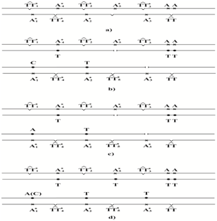
Figure 4 SOS or error-prone DNA synthesis of molecules containing cis-syn cyclobutane thymine dimers in both DNA strands:
a) DNA site containing the cis-syn cyclobutane thymine dimers.
b) Both strands of DNA are synthesized by specialized or modified DNA polymerases, opposite the adenine molecules in rare tautomeric forms A1* cytosine molecule is incorporated.
c) Both strands of DNA are synthesized by specialized or modified DNA polymerases, opposite adenine molecules in rare tautomeric forms A1* adenine molecule is incorporated.
d) Gaps in one nucleotide inserted correctly, opposite adenine molecule in the rare tautomeric form A1* is incorporated molecules of cytosine or adenine
If the Cis-syn cyclobutane thymine dimers are not removed, then this site of DNA is synthesized by DNA polymerases, capable of carrying out the SOS synthesis, or the error-prone synthesis. When there is SOS synthesis of bacteria’s DNA synthesis occurs with the participation of a modified DNA polymerase III E. Coli using the mechanism of "sliding clamp"139 or with the participation of a specialized DNA polymerases IV or VE. Coli.19,20,23,31,33,42‒44 DNA polymerase pol V can proceed synthesis much further from damages.44 If there is translesion synthesis of mammals then DNA polymerases δ or ε of mammals modified by "sliding clamp" the mechanism are involved148‒150 or specialized DNA polymerase, such as Pol η, Pol ζ, Pol κor Pol θof mammals.35,41,143‒147 Let's see what happens when opposite the DNA bases on the site shown in Figure 4a, canonical bases are inserted by specialized or modified DNA polymerases. Cis-syn cyclobutane thymine dimers will result in the targeted mutations.84‒86 In Figure 4, the targeted mutations I have not have represented.
In order to determine which of the canonical bases will be inserted by the modified or specialized DNA polymerase opposite adenines in rare tautomeric forms (Figure 2), consider the constraints on the formation of hydrogen bonds between the bases of the template DNA and the inserted bases. Since the A1* base (Figure 2b) is located in a small neighborhood of the Cis-syn cyclobutane thymine dimer TT4* (Figure 4a), a nucleotide opposite to it can be incorporated by DNA polymerase is able to lead the translesion synthesis, that is, or modified, or specialized DNA polymerase only. DNA polymerases insert canonical nucleotides that can form hydrogen bonds with the matrix bases A1* only. In the case of non-canonical base pairs wrong nucleotide is not deleted.84 The rare tautomer A1* (Figure 2b) cannot form hydrogen bonds with canonical molecule of thymine for steric reasons. But canonical tautomeric forms of cytosine can be incorporated opposite A1* (Figure 5a) and canonical tautomeric forms of adenine can be incorporated opposite A1* (Figure 5b). There is another version of events. However, if the H6'' hydrogen atom in A1* will take the position that the hydrogen atom H6' of molecule of adenine in canonical form takes then adenine A1*' is formed. Molecule of adenine A1*' cannot form hydrogen bonds with any bases in canonical tautomeric forms. Let adenine molecule A2*, which is in rare tautomeric form corresponding to Figure 2c, is located in a small neighborhood of a cyclobutane dimer (Figure 4a). In the synthesis of the DNA site using specialized or modified DNA polymerase A2* adenine molecule can form hydrogen bonds with canonical thymine (Figure 5c). We cannot exclude the fact that the hydrogen atom H6'' of molecule A2* will change its position and will be located as the atom H6' in a canonical molecule of adenine. In this case two hydrogen bonds between adenine A2*' and canonical thymine can form too.
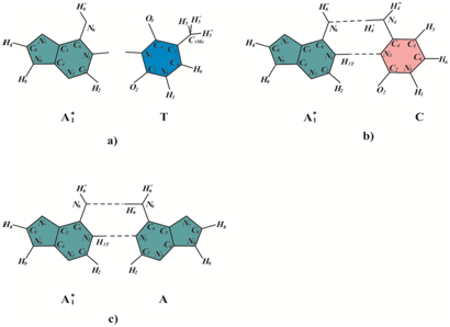
Figure 5 Possible base pairs of adenine molecules in rare tautomeric forms A1* with bases in the canonical tautomeric forms: a) adenine in rare tautomeric form A1* cannot form hydrogen bonds with canonical thymine. b) Pair of adenine molecules A1* with canonical cytosine. c) Pair of adenine molecules A1* with canonical adenine.
Molecules of adenine in rare tautomeric forms A3* (Figure 2d) and A5* (Figure 2f) cannot form hydrogen bonds with any bases in canonical tautomeric forms. Molecule of adenine A4* in rare tautomeric forms (Figure 2e) can form hydrogen bonds with canonical thymine (Figure 5d). Opposite Cis-syn cyclobutane thymine dimers with bases in canonical tautomeric form molecules of the adenine will be in canonical tautomeric form. During DNA synthesis by specialized or modified DNA polymerase inserted canonical bases may form hydrogen bonds with canonical thymines that are part of the Cis-syn cyclobutane thymine dimers. Similarly, opposite of the thymine molecules in canonical tautomeric form that are included in the Cis-syn cyclobutane dimers TT1*, TT2*, TT3*, TT4* and TT5* adenine molecules will be in the canonical tautomeric form.
Let's analyze error-prone and SOS synthesis of the DNA site shown in Figure 4, which takes place by means of specialized or modified DNA polymerases. As we have seen, opposite the adenine A1* DNA polymerases cannot insert canonical thymine so that between them may be formed hydrogen bonds. This means that the canonical base pair and A1* cannot be formed and this inevitably lead to mutations. If cytosine will be inserted the A1*-С pair will be form (Figure 4b). Consequently, in this case, a transition A-Т→G-С appears. But opposite A1* may be inserted molecule of adenine then the pair A1*-A is formed (Figure 4c). (Figure 4b). If opposite A1* canonical adenine will be inserted it would mean the formation of homologous transversion А-Т→Т-А. As is known, some specialized DNA-polymerase, for example DNA polymerase V, provide a high percentage transversions.51
Opposite the molecules adenine A3* and A5* it is impossible to incorporate any of the canonical bases, so that between them and the adenines in rare tautomeric forms hydrogen bonds are formed. The result will be a gaps in a single nucleotide, which is likely, in the future will be inserted correctly. Opposite A2* and A4* can insert the molecule thymine and then the mutations do not appear. Therefore, in this case to untargeted base substitutions mutations can lead only adenine molecules A1*.
The mechanisms of untargeted base substitution mutations formation during error-prone and SOS replication of double-stranded DNA sites containing in both DNA strands molecules of thymine and adenine in rare tautomeric forms closely spaced from cyclobutane pyrimidine dimers
Let us consider a site of DNA, on which in a small neighborhood of cyclobutane pyrimidine dimers with bases in the canonical tautomeric forms pairs base of adenine-thymine in rare tautomeric forms are formed. Let not far from cyclobutane dimers pairs are appeared T1*-A1*, T2*-A2*, T3*-A3*, T4*-A4* andT5*-A5* as it shown in Figure 6a. Bases in rare tautomeric forms are stable. Let's see which the canonical DNA bases can be inserted opposite the molecules adenine and thymine in rare tautomeric forms, illustrated in Figure 2. Structural analysis of the insertion of the bases, made in ref.84 in the study of mechanisms of targeted base substitutions mutations, showed that opposite the molecule of thymine T1* is impossible to incorporate a molecule of adenine in the canonical tautomeric form so that between them hydrogen bonds are formed. But molecules of guanine or thymine can be inserted. Opposite molecule of thymine in rare tautomeric form T2* cannot any canonic bases so that between them hydrogen bonds are formed.84 Opposite the thymine in rare tautomeric forms T3* molecule of the adenine can be inserted.97,98 The rare T5*and T4* tautomers cannot form hydrogen bonds with canonical tautomer of adenine for steric reasons. Canonical tautomeric forms of cytosine and thymine can be incorporated opposite T5*.The rare T4* tautomer is capable of forming hydrogen bonds with cytosine.84 As shown above adenine A1* does not form hydrogen bonds with canonical tautomer of thymine (Figure 5a), but A1* can form hydrogen bonds with canonical tautomer of cytosine (Figure 5b) and with canonical tautomer of adenine (Figure 5c). The rare A3* and A5* tautomers do not form hydrogen bonds with canonical tautomers of DNA bases. The rare A2* and A4* tautomers of adenine can form hydrogen bonds with thymine.
Let us the site of DNA shown in Figure 6a is synthesized as a result of error-prone or SOS synthesis. This means that, firstly, these damage of DNA are not removed, and secondly, the site will be inserted by specialized or modified DNA polymerases. Canonical tautomeric forms of guanine or thymine can be incorporated opposite T1*.In this case, A-Т→G-С transition or А-Т→Т-А homologous transversion will result (Figure 6b). Thus, if the near cyclobutane dimers the rare T1* tautomer is formed and, if it proved to be stable, then the error-prone or SOS DNA synthesis, it will inevitably lead to untargeted base substitution mutations, transitions or homologous transversions. But the rare T1* tautomer cannot result in A-Т→С-Gtransversions.
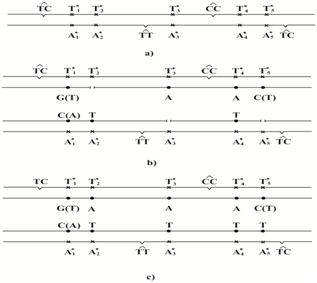
Figure 6 SOS or error-prone synthesis of DNA molecules containing the base pairs adenine - thymine in rare tautomeric forms in the small vicinities of any cyclobutane dimers:
a) Site of DNA containing molecules of adenine and thymine in rare tautomeric forms in the small vicinities of any cyclobutane dimers.
b) Both strands of DNA are synthesized by specialized or modified DNA polymerases; opposite the molecules of adenine in rare tautomeric form A1* are inserted canonic molecule of cytosine or adenine. opposite the molecules of thymine in rare tautomeric form T1* are inserted canonic molecule of guanine or thymine; opposite the molecules of thymine in rare tautomeric form T5* are inserted canonic molecule of cytosine or thymine.
c) Gaps in one nucleotide are inserted correctly; opposite the A1* are inserted canonic molecule of cytosine or adenine; opposite the T1* are inserted canonic molecule of guanine or thymine. Opposite the T5* are inserted canonic molecule of cytosine or thymine.
The rare T2* tautomer cannot form hydrogen bonds with any canonic tautomers of DNA bases for steric reasons. Therefore, likely DNA synthesis will result in a one-nucleotide gap (Figure 6b), which will be further incorporated by unmistakable manner (Figure 6c). Since opposite the thymine in rare tautomeric forms T3* molecule of adenine can be inserted, it is likely it will not result in mutations. Molecules of the thymine in rare tautomeric form T4* may result in transversion A-Т→С-G only. Rare T5* tautomer is possible to incorporate molecules of cytosine or thymine84 they can result in transversion A-Т→С-G or homologous transversions А-Т→Т-А only (Figure 6b). Just like in the previous case, A1* can result in A-Т→G-С or to А-Т→Т-А homologous transversion. A one-nucleotide gaps are formed opposite molecule of adenine A1*', A3* and A5* is likely in the future they will be inserted correctly. Molecule of thymine can be inserted opposite A2* and A4*, they may not result in mutations.
As we can see, from all possible rare tautomeric forms of adenine and thymine, only three, namely, T1*, T4*, T5* and A1*, may cause untargeted base substitution mutations. Thus molecules of thymine T1* and adenine A1* in rare tautomeric form may result in transitions A-Т→G-С. Molecules of the thymine T1*, T5* and adenine A1* in rare tautomeric forms can lead to homologous transversions А-Т→Т-А, and molecules of the thymine T5* and T4* in rare tautomeric form may result in transversion A-Т→С-G. Thus, a rough estimate predicts that during the untargeted base substitution mutations formation which are formed during the formation of stable rare tautomeric forms of thymine and adenine, there is a 20% of transitions and 80% of transversions. As is known, untargeted mutagenesis is characterized by a high percentage of education transversions.34,36,66
There is currently no complete understanding of the question of the nature and mechanisms of formation of untargeted bases substitution mutations. Mechanisms of various mutations are usually considered as part of the polymerase model of mutagenesis. I suggested, and developing alternative polymerase-tautomeric models of ultraviolet mutagenesis, bystander effects and genomic instability. In this paper, it is developed mechanisms of untargeted base substitutions mutations induced by molecules of thymine and adenine in rare tautomeric forms. Untargeted bases substitution mutations are substitution mutations that appear on the so-called not damaged DNA sites. Untargeted base substitution mutations can be caused, for example, irradiation with ultraviolet light of DNA molecules. As a result, as is known, cyclobutane pyrimidine dimers are formed. Ultraviolet radiation can lead to change in tautomeric states of DNA bases. Molecules of thymine and adenine can form five rare tautomeric forms. These rare tautomeric form will be stable if the corresponding nucleotides are part of cyclobutane dimers or are in a small neighborhood of them. If the tautomeric states in a pair of DNA bases are changed its tautomeric states change in both DNA bases. It changes in thymine, which is part of a cyclobutane dimer and adenine to which they are linked by hydrogen bonds.
Ii is considered a site of DNA, on which Cis-syn cyclobutane thymine dimers with bases in rare tautomeric forms appeared in both strands of DNA close to each other. Besides it is considered a site of DNA, on which in a small neighborhood of cyclobutane pyrimidine dimers with bases in the canonical tautomeric forms pairs base of adenine-thymine in rare tautomeric forms are formed. These sites are synthesized as a result of error-prone or SOS synthesis. Structural analysis indicates that canonical tautomeric forms of thymine cannot be incorporated opposite A1*. But canonical tautomeric forms of cytosine or adenine can be incorporated opposite A1*. Rare A1* tautomer of adenine may result in a untargeted transition A-Т→G-С or a untargeted homologous transversion А-Т→Т-А. Molecule of thymine can be inserted opposite A2*and A4*; molecule of adenine can be inserted opposite T3*; it is likely they will not result in mutations. The rare A3*, A5* and T2* tautomers do not form hydrogen bonds with any canonical tautomers of DNA bases. So they cannot result in the base substitution mutations. Rare T1* tautomer of thymine may result in A-Т→G-Сuntargeted transition or А-Т→Т-Аuntargeted homologous transversion. Molecules of the thymine in rare tautomeric form T4* may result in transversion A-Т→С-G only. Rare T5* tautomer can result in transversion A-Т→С-G or homologous transversions А-Т→Т-А.
Thus, it is shown that the relationship between the type of primary DNA damage and the resulting kinds of mutations is not always simple. The same potential mutation can lead to transition or transversions. Which the mutation is formed depends on kind of rare tautomeric form of corresponding bases and a specialized DNA polymerase which is involved in DNA synthesis. The molecule of thymines T1*, T4*, T5* and of adenine A1* located near the cyclobutane pyrimidine dimers may cause untargeted base substitution mutations. This conclusion is reached taking into account the fact that there is a control of the side groups of bases in the synthesis of DNA.154 If the mutagenic load is such that such control does not work, then it may appear Hoogsteen155,156 base pairs. Polymerase-tautomeric model predicts that under the formation of untargeted base substitution mutations 20% of transitions and 80% of transversions are formed. As is known, untargeted mutagenesis is characterized by a high percentage of education transversions.
The term untargeted mutations suggest that these mutations are formed on undamaged DNA sites. As it is have shown in this paper, this is not right. It should be assumed that untargeted mutations are a mutations appearing on DNA damages unable to stop the synthesis of DNA. This hypothesis was tested by biological methods only. Firmly established facts show that the so-called untargeted mutations appear on DNA sites, in which, using biological methods, no DNA damages was found. This does not mean that using other methods such as thermally stimulated luminescence, such DNA damages are not to be found. It is clear that in order to explain the nature of untargeted base substitutions mutations have not the slightest need to involve the ideas used now to explain bystander effects. They are easily and naturally explained by polymerase-tautomeric model.
Some substances, for example, xenobiotics, promote oxidative stress with the release of free radicals.157 Free radicals can change the tautomeric forms of DNA bases by exactly the same mechanism as ultraviolet irradiation of DNA. This can lead to targeted and untargeted mutagenesis, as well as to the instability of the genome. Therefore, the polymerase-tautomeric model is able to explain the mechanisms formation of targeted base substitution mutations, targeted insertions, targeted deletions, targeted complex insertion, targeted base substitution mutations and hot and cold spots of UV-induced mutagenesis. The polymerase-tautomeric model for bystander effects is able to explain the mechanisms formation for delayed targeted base substitution mutations and untargeted base substitution mutations.
None.
Author declares that there is no conflict of interest.

©2017 Grebneva. This is an open access article distributed under the terms of the, which permits unrestricted use, distribution, and build upon your work non-commercially.