International Journal of
eISSN: 2573-2838


Research Article Volume 4 Issue 4
1ExoAtlet LLC, Russia
2Institute of Mechanics of Moscow State University, Russia
3Institute of phtisiopulmonology, Russia
Correspondence: Pismennaya EV, Institute of Mechanics of Moscow State University, , Tel +7 (495) 374-85-30
Received: August 24, 2018 | Published: August 29, 2018
Citation: Pismennaya EV, Petrushanskaya KA, Shapkova EY. Formation of the locomotor stereotype in patient with consequences of the spinal cord injury (single case). Int J Biosen Bioelectron. 2018;4(4):209-215. DOI: 10.15406/ijbsbe.2018.04.00128
The process of emergence of the locomotor act under the influence of training in the exoskeleton in combination with transcutaneous electrical stimulation of the spinal cord has been illustrated in this article on the concrete example of patient with the spinal cord injury. It has been revealed, that not simply improvement of walking takes place in the process of many-days training, but its appearance. Authors considered the stages of formation of the locomotor act. They revealed the distinctive features of the first steps of patient, appearance of the locomotor act in the process of the first training. Authors retraced this process from the absence of real walking (ambulation in the walker with the attached step to emergence of the pathological walking, then development of phenomena, peculiar to the pathological walking, and at last – formation of more right stereotype of walking. Authors demonstrated, that the following perturbations take place during walking in the exoskeleton: increase of the support area of the feet and of the extreme values of the ground reaction force, disappearance of the phenomena of the dynamic and temporal asymmetry, increase of duration of the double-support phase, what causes improvement of the biomechanical picture of walking after the session and emergence of the walking, put pathological one. The following stage is accompanied by development of the elements of the pathological walking, namely of change of correlation of the stance and swing phase in favour of the stance phase, further increase of duration of the double-support phase, expressed asymmetry of all parameters of walking, presence of the pathological and compensatory mechanisms and, at last, formation of more right stereotype of walking, corresponding with growth of walking velocity and double step length.
Keywords: rehabilitation, exoskeleton, spinal cord injury, locomotor system, biomechanical structure of walking, vertical component of the ground reaction force
The problem of rehabilitation of patients with diseases of the locomotor system has become one of the most actual in the recent years. Nowadays this problem becomes extremely important in such diseases as multiple sclerosis, stroke, infantile cerebral palsy and consequences of the spinal cord injury. This phenomenon is connected with the fact, that number of patients with such diseases has a tendency to growth, rehabilitation of these patients is rather expensive, and at last, these patients often need constant and many-sided help from the relatives.1–10 Nowadays acute necessity for search for the most effective methods and means of rehabilitation of such patients appeared in Russia. The main purpose of these methods is to receive the best restorative effect for the shortest time. Application of the exoskeleton during walking of patients with different diseases of the locomotor system is one of such methods. Medical exoskeleton “ExoAtlet”, elaborated in Russia, is intended for assistance to people with the limited physical possibilities. This exoskeleton may be applied both for the medical and for the social rehabilitation of patients with the locomotor disturbances. As it has already been mentioned, spinal cord injury is one of the most serious disease. Traumatic injury of the spinal cord is a reason of the remarkable changes of the person’s life. This is true not only for the main physiological processes (disorder of the motional functions, of the function of the pelvic organs, of the respiratory and cardio-vascular systems, trophic disorders), but essentially changes quality of life of patient and his family, demands adaptation to quite new social, economical and professional conditions of existence. 2–5,8 Different authors note, that steady growth of the part of the spinal cord injury in the structure of the combined trauma is observed in Russia.2–8 According to data of M.A. Leontiev, for the last 70 years number of patients with the spinal cord injury increased in 200 times.7,8 Every year 8 thousand people receive spinal cord injury in Russia, and 10 thousand - in USA. Trauma of the spinal cord affects two or more levels in 10-12% of cases, multiple injuries are observed in 34% of patients.8 Severity of this process is conditioned by the character, length, level and the degree of injury of the spinal cord.1–10 But even inconsiderable injury of the spinal cord dooms majority of patients to many-years sufferings.
That’s why it becomes necessary to give constant and many-sided help to such people. Investigations, carried out in Pirogov National medico-surgical centre, showed, that the following changes take place after two-weeks training in the exoskeleton in patients with the spinal cord injury:
The next problem was to determine the most effective combination of training in the exoskeleton with the other methods. From our point of view, combination of walking in the exoskeleton with the functional electrical stimulation (FES) of muscles or with the stimulation of the spinal cord present the best variants for rehabilitation of such patients.13,14 The purpose of this work was to evaluate the results of the course of rehabilitation by means of combination of walking in the exoskeleton with transcutaneous electrical stimulation of the spinal cord by the example of patient with the spinal cord injury.
By means of the force-measure insoles the following parameters of walking have been investigated: main indices (double step length, duration of the locomotor cycle, walking velocity, cadence), temporal indices (duration of the stance and swing phases, of the double-support phase, T-interval, rhythm coefficient), dynamic indices (vertical component Rz of the ground reaction force).15–17 Choice of this method for investigation of the biomechanical parameters of walking is explained by the following reasons.
From our point of view, it is expedient to consider the results of the course of rehabilitation by means of combination of walking in the exoskeleton with transcutaneous electrical stimulation of the spinal cord by the concrete example. Patient Sablin E.N. Age – 38. Diagnosis: consequences of the spinal cord injury, fracture of Th12, fracture-dislocation of L1. Severity of the disease is estimated as type C according to Frankel scale. Period after the spinal cord injury comprised 2,5 years. The level of mobility was equal to 4 points according to Rivermid scale. All trainings and all investigations have been carried out on the base of the Sankt-Petersburg institute of Phtisiopulmonology.
This study was carried out in accordance with the recommendations of the Institute Guidelines for Clinical Trials, Institutional Review Board of the Sankt-Petersburg institute of Phtisiopulmonology. Patient gave the written informative consent in accordance with Declaration of Helsinki. The protocol was approved by the Institutional Review Board of Moscow Regional Research Clinical Institute. The Institute Guidelines for Clinical Trials conforms to the ethical principles of the Declaration of Helsinki. The course of rehabilitation consisted of 12 trainings of walking in the exoskeleton. At the beginning of the course patient needed help of two assistants. After 3 sessions patient could already walk with the support on the Canadian canes with one assistant. Trainings in the exoskeleton were accompanied by tonic low-frequency noninvasive electro stimulation of the middle part of the lumbar enlargement of the spinal cord with displacement of electrodes on the skin (-) – above the vertebrae Th12, (+) – on the anterior abdominal wall, according to method of E. J. Shapkova.18 Stimulation was realized by means of the portable electro stimulator. Duration of the session comprised 30 minutes. During walking in the exoskeleton cadence was equal to 40 steps/vin, double step length – 0.6 m, and mean walking velocity – 0,2 m/s, for all this every session patient made 1200 steps and overcame a distance of 360 m.
Biomechanical characteristic of walking
Walking of this patient is interesting because it is possible to retrace its peculiarities from the moment, when patient was able to make his first steps with the help of walker till the moment, when he could walk independently along the horizontal surface with two Canadian canes. Before the course of rehabilitation patient could make only 2- 4 steps. Patient moves very slowly with the support on the walker, with the attached step, for all this he doesn’t practically lean on the right leg, practically completely «hungs» in the walker, during the heel-support of the left leg patient very quickly shifts the walker (Figure 1). Biomechanical parameters have been investigated in four variants:

Figure 1 Cyclogram of the locomotor cycle of patient S-n. Dyagnosis – consequences of the spinal cord injury, fracture of Th12, fracture-dislocation of L1.
Parameter |
Norm, n=10 |
Patient with SPI |
|||||
|---|---|---|---|---|---|---|---|
Before the course |
% to norm |
After the session |
% to the init. data |
After the course |
% to the init. data |
||
Double |
1.42 |
0.3 |
21 |
0.5 |
167 |
0.62 |
207 |
Duration of the |
1.21 |
2.31 |
191 |
2.3 |
100 |
2.5 |
108 |
Walking |
1.17 |
0.13 |
11 |
0.22 |
169 |
0.25 |
192 |
Walking |
99 |
52 |
53 |
52 |
100 |
48 |
92 |
Table 1 Main parameters of walking in norm and in patient with the SPI S-n
Parameters |
Norm |
Before the course |
After the session |
After the course |
||||||||||
|---|---|---|---|---|---|---|---|---|---|---|---|---|---|---|
L |
R |
L |
R |
% to norm |
L |
R |
% to the init. data |
L |
R |
% to the init. data |
|
|||
L |
R |
|
|
L |
R |
|
|
L |
L |
|||||
Duration of the stance phase (%) |
62.4 |
62.6 |
69.5 |
44.3 |
111 |
71 |
73.3 |
70.0 |
105 |
158 |
68.5 |
69.4 |
99 |
157 |
Duration of the swing phase (%) |
37.6 |
37.4 |
30.5 |
55.7 |
81 |
149 |
26.7 |
30.0 |
88 |
54 |
31.5 |
30.6 |
103 |
55 |
Duration of the double-support phase (%) |
12.7 |
12.3 |
3.2 |
10.6 |
25 |
86 |
7.3 |
36.0 |
228 |
340 |
11.1 |
26.8 |
347 |
253 |
Rhythm coefficient |
0.99 |
0.55 |
|
0.89 |
|
0.97 |
|
|||||||
Table 2 Temporal parameters of walking of patient S-n before and after the course of training in the exoskeleton
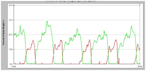
The main parameters of walking in norm and in patient S-n are presented in Table 1. As may be seen from this table, mean walking velocity is reduced in 9 times in comparison with norm 9 (0.13 m/s) at the account of shortening of the double step length in 4,7 times (0.3 m) and reduction of the walking cadence – in 1.9 times (52 steps/min). Temporal indices of walking change to a greater extent in this patient (Table 2). In comparison with norm growth of duration of the stance phase (69,5%) and diminution of duration of the swing phase (30,5%) is observed in the left lower extremity. At the same time the contrary picture is seen in the right leg, namely considerable decrease of duration of the stance phase (44,3%) and increase of duration of the swing phase (55,7%), what causes remarkable diminution of the rhythm coefficient. The essential decrease of duration of the double-support phase takes place in both legs, in particular – in the left leg – by a factor of 4 (3/2%), and in the right leg – by 15% (10,6%), what points to absence of the static stability during walking. The dynamic parameters of walking are remarkably transformed during walking of patient before the course of rehabilitation without exoskeleton (Figure 2). The pronounced asymmetry of the dynamic parameters of walking is also retraced here. The vertical component of the ground reaction force in the left leg acquires the evident triangular form. The extreme value of 33% is developed only by 30% of the locomotor cycle. All extreme values are remarkably reduced in the right leg. Nevertheless Rz curve has a right form, approaching to two-peaked. The extreme values of the vertical component Rz of the right leg comprise 16,3% - for heel-strike, 13.2% - for the minimal value, 20.6% – for push-off (Table 3). So, the severe disturbance of locomotion takes place in patient S-n in comparison with norm, namely remarkable reduction of mean walking velocity, cadence and double step length, factual absence of the static stability during walking (considerable decrease of duration of the double-support phase in both legs), change of the form of the vertical component of the ground reaction force from two-humped to the triangular one with one maximal value in the left leg, diminution of the extreme values of the vertical component of the ground reaction force in both lower extremities.
Parameters, % of BW |
Norm |
Before the course |
After one session |
After the course |
||||||||||
|---|---|---|---|---|---|---|---|---|---|---|---|---|---|---|
L |
R |
L |
R |
% to norm |
L |
R |
% to the init. data |
L |
R |
% to the init. data |
||||
L |
R |
L |
R |
L |
L |
|||||||||
Heel-strike, % |
118.5 |
118.5 |
– |
16.3 |
– |
14 |
– |
19.6 |
– |
120 |
76.6 |
46.4 |
– |
284 |
Minimum, % |
72.0 |
72.0 |
– |
13.2 |
– |
18 |
– |
17.9 |
– |
136 |
64.5 |
35.2 |
– |
267 |
Push-off, % |
125.7 |
125.7 |
33.1 |
20.6 |
26 |
16 |
33.9 |
21.1 |
102 |
102 |
84.2 |
50.3 |
254 |
244 |
Table 3 Dynamic parameters of walking of patient S-n before and after the course of training in the exoskeleton

Figure 2 Ground reaction force in percent to body weight during walking without exoskeleton before the course of training. Red line – right leg, green line- left leg. Along the abscises axis – time in seconds.
Biomechanical parameters of walking in the exoskeleton
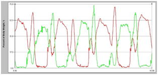
During training in the exoskeleton the walking stereotype, peculiar to a healthy man at a slow cadence, is imposed to this patient.19 For all this main and temporal parameters of walking in the exoskeleton don’t essentially change in comparison with the corresponding parameters of walking of the healthy people in the exoskeleton.14,19 Nevertheless evident growth of duration of the double-support phase is examined in both legs in patient, what may be explained by prolonged transfer of crutches and support on them in this phase. Considerable improvement of the extreme values of the vertical component Rz of ground reaction force takes place during walking in the exoskeleton. It is possible to see even distribution of the ground reaction force on both of the lower extremities. Increase of the value of the heel-strike and push-off is examined already in the beginning of the course. For all this Rz curve acquires the evident two-humped form in both lower extremities (Figure 3). Dynamic asymmetry practically disappears during walking in the exoskeleton. So, value of heel-strike comprises 46% in the left leg, and 51,8% - in the right leg, value of the minimum comprises correspondingly – 27,2% and 30.7%, of the push-off - 52,8% and 49,4%. Consequently, during walking in the exoskeleton amplitude of both pushes considerably rises simultaneously with elimination of the temporal and dynamic asymmetry.

Figure 3 Ground reaction force in percent to body weight during walking of patient in the exoskeleton in the beginning of the course.
Results after the session of training
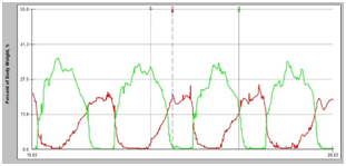
Results showed that already the next day after the session in the very beginning of the course essential improvement of the main and temporal indices of walking is seen in this patient. In particular, double step length increases from 0.3 to 0.5 m, walking velocity - from 0.13 m/s to 0.22 m/s, while walking cadence remains unchanged. Nevertheless the temporal structure of walking is essentially transformed. First of all, greater symmetry of the temporal parameters of walking is observed in a day after the session. In particular, duration of the stance phase is equal to 73,4% in the left leg, and 68.5% – in the right leg, and of the swing phase correspondingly – 26,6% and 31,5%. It is necessary to note, that changes of the temporal structure of walking in the left lower extremity are inconsiderable and mostly concern growth of duration of the double support phase. Duration of the stance and double-support phases remarkably increases in the right leg, while duration of the swing phase tends to diminish. It is of interest, that in the beginning of the course only slight growth of the extreme values of the vertical component of the ground reaction force takes place, in particular, of the heel-strike in the right leg (Figure 4). So, the certain changes are observed after the first training in the exoskeleton, which concern predominantly the temporal asymmetry, but don’t practically influence on the dynamic parameters of walking Figure 6. The averaged graphs of the vertical component of the ground reaction force in percent to body weight before and after the course of training in the exoskeleton. Black arrows mark ground reaction force before the course, red arrows mark ground reaction force after the course. As may be seen from these graphs, after the course of training in the exoskeleton the extreme values of Rz curve increased by a factor of 2, and, besides, growth of duration of the stance phase in the right leg also took place.

Figure 4 Ground reaction forces in percent to body weight the next day after walking in the exoskeleton.

Figure 5 Ground reaction force in percent to body weight during walking without exoskeleton after the course of training in the exoskeleton.

Figure 6 The averaged graphs of the vertical component of the ground reaction force in percent to body weight before and after the course of training in the exoskeleton.
Results after the course of training
As evident from Table 1, essential improvement of the main parameters of walking is observed after the course of complex rehabilitation, in particular, growth of walking velocity in 2 times, and this growth of velocity is realized exclusively at the expense of increase of step length (from 0,3 to 0,62 m), while cadence tends to diminish from 53 to 48 steps/min. Duration of the stance and swing phases is approximately the same in both legs and comprises correspondingly 69% - for the stance phase and 31% - for the swing phase. Temporal asymmetry is noted only in duration of the double-support phase. Evident rise of the double-support phase takes place in comparison with the beginning of the course, what points to appearance of the static stability and greater supportability of the lower extremities. Transformation of the dynamic parameters of walking after the course of training also confirms growth of the support and push functions of the lower extremities (Figure 5) (Figure 6). In particular, after the course of training considerable increase of the extreme values of the vertical component of the ground reaction force is noted. Besides, now patient is able to support not only on the heel, but also on the lateral part of the foot, but he can’t yet support on the metatarsal zone of the foot. So, remarkable improvement of the locomotor function is observed only after two-weeks training of walking in the exoskeleton, which is revealed in increase of walking velocity and step length, in complete disappearance of the temporal asymmetry, in growth of stability, in change of the form of the vertical component of the ground reaction force from the triangular to two-humped, in rise of the extreme values of the ground reaction force, in widening of the support area of the foot from the heel to the lateral part of the foot, in lesser application of the additional support.
In spite of the fact, that we applied many times term ”walking” to the phenomenon, which we observed in the beginning of the course, such ambulation can’t be called walking in the classical sense of this term. Such ambulation can’t even be called pathological walking because of some reasons.19 Firstly, according to many years investigations, walking cadence is diminished in all types of pathological locomotion, otherwise duration of the locomotor cycle tends to grow. As pathology becomes more severe, duration of the stance phase more and more increases, while duration of the swing phase more and more decreases. In the most severe cases in patients with the spinal cord injury duration of the stance phase achieves 90-92%, and duration of the swing phase is equal to 8-10%. Secondly, provision of stability is achieved by change of correlation of duration of the stance and swing phase in favour of the stance in the side of the intact extremity (or less affected lower extremity). Thirdly, as the pathology becomes more and more serious, duration of the double-support phase must also grow more and more, in the most severe cases it achieves 40-42%, what also promotes increase of the static stability during walking.19,20 In this case duration of the swing phase in the right leg is greater in comparison with the stance phase, what indicates, that patient hasn’t been trained the right skill of walking yet. Double-support phase is practically absent in this patient, especially in the left leg and this phase is remarkably low in the right leg, what points to the essential instability during walking. Probably, this fact is connected with growth of duration of the swing phase on the right leg, because most part of the locomotor cycle the right extremity is situated above the support, what also causes considerable instability during locomotion. So, it is believed, that in the stage of formation of the walking stereotype, i.e. in the moment of transition from absence of walking to the first steps with the walker it is a possible to examine phenomenon of so called «kangaroo step”, what is accompanied by increase of duration of the swing phase and diminution of duration of the stance phase and especially of the double-support phase of one leg.4 Consequently, very great instability is noticed during walking, because extremity is above the support surface most part of the locomotor cycle. Biomechanical picture of walking, typical of the pathological one, begins forming just after the first session. It is necessary to note, that walking velocity increases the next day after the first session from 0,13 m/s to 0.22 m/s, i.e. after the first session patient continues walking with the velocity, imposed him by the exoskeleton. Nevertheless support and push function don’t grow in the first stage. At the next stage in the process of the further training the traditional picture of the pathological gait is being developed. This picture is revealed in the following peculiarities: in remarkable growth of duration of the stance phase and reduction of duration of the swing phase, in presence of asymmetry, in absence of the toe-off support and, consequently, T-interval (time from heel-off of one leg till the beginning of the stance phase of another leg), in change of the form of the vertical component of the ground reaction force from two-peaked to the trapezium, in considerable displacement of all extreme values of the ground reaction force to the right along the temporal axis. At last, at the third stage it becomes possible to achieve the right temporal structure of walking and right distribution of loading, approximately equal duration of the stance and swing phases, otherwise to overcome temporal asymmetry. And then in the next stage growth of the extreme values of the vertical component Rz of the ground reaction force takes place.
It is believed, that until the sufficient supportability of the lower extremity is not achieved, until normal stepping on the support is not provided, until loss of the necessary information from the foot takes place, it is factually impossible to achieve increase of the support and especially push function of the lower extremities. The unique experiment of A.S. Vitenson with anesthesia of the distal part of the lower extremities demonstrated, that afferentation from the distal part of the lower extremity is lost in anesthesia of the foot and ankle joint (AJ).21 Anesthesia of the foot and AJ eliminates the great receptor field of the foot, formed by the skin receptors, articular bursas, ligaments, tendons and short muscles of the foot. From this number of receptors only the joint receptors give signals of inter location of the extremity with high accuracy, and the proprioceptive elements, besides, transfer high-differentiated information of all interactions of the person with the support surface during standing and walking.22 Afferent fibers of all above-mentioned receptors comprise the group of so called receptors of the flexor reflex (AFR). Signals from periphery, incoming to the spinal cord along the AFR, take part in formation of the intra spinal program of movements in animals with the intact afferent entrance.23 Probably, training in the exoskeleton promotes remarkable increase of the receptive field of the foot, formed by the afferents of the flexor reflex, what points to the fact, that activity of muscles in person is realized with participation of the afferents, informing the central nervous system of position of the segments, forming the joint about direction and velocity of their displacement. So, only in two weeks it is possible to retrace the process of transformation from the factual absence of walking at first to appearance of the pathological walking, then to development of all phenomena, peculiar to pathological walking (change of correlation of duration of the stance and swing phases in both lower extremities, asymmetry of all parameters of walking, growth of the static stability, presence of pathological and compensatory mechanisms) and at last to improvement of the process of locomotion and increase of walking velocity. In 1 year after the course of rehabilitation patient refused to apply a wheel-chair. At present patient walk with the support on two Canadian canes.
None.
The author declares that the research was conducted in the absence of any commercial or financial relationships that could be construed as a potential conflict of interest.

©2018 Pismennaya, et al. This is an open access article distributed under the terms of the, which permits unrestricted use, distribution, and build upon your work non-commercially.