eISSN: 2471-0016


Case Report Volume 1 Issue 2
1Department of Pathology, Kathmandu University School of Medical Sciences, Nepal
1Department of Pathology, Kathmandu University School of Medical Sciences, Nepal
Correspondence: Rachana Dhakal, Department of Pathology, Kathmandu University School of Medical Sciences, Nepal
Received: January 01, 1971 | Published: September 9, 2015
Citation: Dhakal R, Lakshmi R, Sumeet D. Malignant eccrine spiradenoma with squamoid and chondroid differentiation. Int Clin Pathol J. 2015;1(2):35–36. DOI: 10.15406/icpjl.2015.01.00008
Malignant eccrine spiradenoma (MES) is an extremely rare sweat gland tumor that arises in a pre-existing benign eccrine spiradenoma. We describe a case of malignant eccrine spiradenoma of a 41-year-old male with a history of swelling over nape of neck for 6yrs and recent onset of increase in size since 6 months. Microscopically, the tumour showed benign component, which was composed of two nodules with interwining bands of 2types of epithelium-, inner differentiated cuboidal cells and peripheral undifferentiated cells. Malignant component was composed of large clear cells with areas of Squamoid and Chondroid differentiation, lymph nodes showed metastasis. With all these features, a diagnosis of malignant eccrine spiradenoma with Squamoid and Chondroid differentiation was made.
Keywords: malignant eccrine spiradenoma, skin tumour, differentiation
Malignant eccrine spiradenoma (MES) is an extremely rare sweat gland tumor. It may develop de novo(malignant eccrine poroma or mucinous carcinoma) or arise in pre-existing benign eccrine spiradenoma.1 However most tumors are presented in the latter mode. It usually presents as a small, firm, reddish painful and small solitary nodule.2,3 Etiology is unknown although previous trauma is believed to be an implicated factor.1,4 In this report, we present an additional case of malignant eccrine spiradenoma diagnosed on 41 year old male which arises in a very unusual location with a peculiar histopathology.
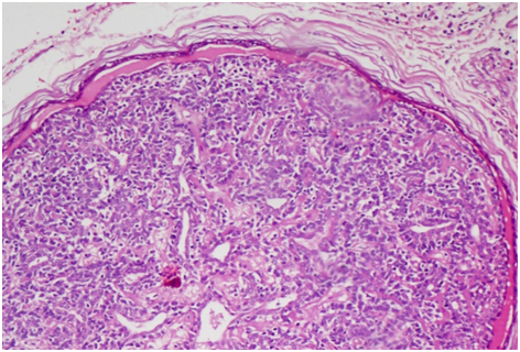
A 41years male patient, presented with a history of swelling over nape of neck for 6 yrs and recent onset of increase in size since 6 months. Patient underwent complete excision. Grossly, mass appeared grey white measuring 1.7cm in diameter. On microscopy, nodules of benign adnexal tumour undergoing malignant transformation (Figure 1) were noted. Benign component is composed of 2 nodules with interwining bands of 2 types of epithelium- inner differentiated cuboidal cells and peripheral undifferentiated cells (Figure 2 & Figure 3). Malignant component was composed of large clear cells with areas of Squamoid (Figure 4) and Chondroid (Figure 5) differentiation, lymph nodes showed metastasis. With all these features, a diagnosis of malignant eccrine spiradenoma with Squamoid and Chondroid differentiation was made.

In 1972 Dabska reported the first case of malignant transformation of eccrine spiradenoma, which is an extremely rare tumor that arises from preexisting eccrine spiradenoma.5 It is an uncommon tumor that occurs in young adult without any sex predilection. The tumor usually presents as a solitary firm round dermal nodule on any part of the body but most frequently on face, scalp, trunk and proximal parts of limbs.6 The average size of malignant eccrine spiradenoma at presentation is 3.9cm (range 0.5-15cm).6 The malignant eccrine spiradenoma almost always arises from a preexisting benign eccrine spiradenoma after a variable latent period, which may be as long as 75 years.7 It generally begets medical attention when a pre-existing undiagnosed lesion rapidly enlarges, changes color, ulcerates, or becomes painful and tender.8 Malignant eccrine spiradenoma metastasizes to regional lymph nodes, lungs, brain, and liver (in descending order of frequency).6 Management depends on proper diagnosis which is based on histopathologically findings. Histopathologically, proliferation of cells with hyper chromatic nuclei, increased mitoses, loss of Periodic Acid-Schiff positive basement membrane cords and invasion of the surrounding tissues characterize malignant transformation in eccrine spiradenoma. It shows focal squamous differentiation, which may be florid in rare instances. Less frequently, areas of spiradenoma near or in transition with a malignant tumor such as rhabdomyosarcoma, osteosarcoma, leiomyosarcoma, and chondrosarcoma may be present.9,10 Immunohistochemical, malignant eccrine spiradenoma exhibit variable expression of cytokeratins, carcinoembryonic antigen, epithelial membrane antigen and S100 protein. Over expression of p53 protein in benign spiradenoma has been associated with malignant transformation, usually into a carcinoma. However, carcinosarcomatous transformation has also been reported.11 Radiation therapy has little role in the management of sweat gland tumors.12 These lesions are rare so the experience with chemotherapy is limited.7 Symptomatic improvement and shrinkage of the tumor with tamoxifen therapy in a patient with estrogen receptor positive eccrine adenocarcinoma has also been reported.6 Recurrences after treatment are frequent and often occur after incomplete tumor excisions. Hence, aggressive surgical treatment must be performed.
None.
The author declares no conflict of interest.

©2015 Dhakal, et al. This is an open access article distributed under the terms of the, which permits unrestricted use, distribution, and build upon your work non-commercially.