eISSN: 2471-0016


Research Article Volume 4 Issue 1
1Department of Urology, Nephrology and Andrology of Kharkov National Medical University, Ukraine
1Department of Urology, Nephrology and Andrology of Kharkov National Medical University, Ukraine
Correspondence: Knigavko Oleksandr, Associate Professor, Department of Urology, Nephrology and Andrology of Kharkov National Medical University, Head of Laboratory of Andrology and Human Reproduction, Ukraine, Tel 3805 0401 2543
Received: January 01, 1971 | Published: January 9, 2017
Citation: Andriy A, Oleksandr K. Histological examination of graft for best choice of corporoplasic. Int Clin Pathol J. 2017;4(1):17–20. DOI: 10.15406/icpjl.2017.04.00083
Treatment of penile deviations with surgical method is still a big problem in view of the frequent complications. During 2006-2016 we performed immunohistological analysis of 168 grafts for patients with penile deviation. Next, auto grafts-fascia lata femoris (FLF), vena femoralis (VF), tunica vaginalis testis (TVT), as well as xenograft-lyophilized bovine pericardium (LBP) were investigated. Recommendations considering the use of grafts cause dpenile deviations were done.
Keywords: erectile dysfunction, penile deviations, corporoplasty, peyronie's disease, grafts
FLF, fascia lata femoris; VF, vena femoralis; LBP, lyophilized bovine pericardium; TVT, tunica vaginalis testis; MCAB, murine monoclonal antibody
The number of patients that are seeking for adequate surgical treatment for both congenital and acquired penile deviations has increased recently. Patient's appeals are associated not only with the presence of a deviation that causes aesthetic dissatisfaction and technical difficulties during coitus, but with concomitant erectile dysfunction, according to 25% -80% of cases.1 Normal flexibility and extensibility of the penile tissues are significant for an erection. Symmetrical disturbance of the tension during erection leads to a curvature of the penis. Congenital curvature is connected with:
On the issue of expediency of surgical correction of the congenital and acquired deformities of the penis, disagreement in the medical community does not arise. The debate continues on the problem of finding the best ways of the surgical correction to save a length and a erectile function of a penis. Despite significant progress in penile surgery, unsatisfactory results of surgical treatment of the penile deviations still remain at a high level (11-70% of cases).2 Dissatisfaction with the results of surgical treatment in the case of Peyronie's disease, and isolated congenital erectile deformation is between 15% and 63%.3 The main reason for postoperative dissatisfaction is recurrence of the deviation and the addition or increase of existing erectile disorders. It has been noted, that if a corporoplasty with grafts is made, the structure of the graft and the structure of the tunica albuginea can be not the same that in postoperative term can cause or exacerbate pre-existing erectile dysfunction due to disorder of arterial inflow or increased venous outflow or infringement of both the mechanisms.4 Nowadays, there is no ideal surgical technique and the ideal graft in the treatment of penile deviations.5
Although the percentage of unsatisfactory results of corporoplasty with grafts, a comparative morphological analysis of the advantages and disadvantages of using auto- and xenografts in the surgical treatment of penile deviations is still not carried out.
Aim
Aim of the study selection of “ideal” graft for different form of penile deviations based on immune histological investigations of plaque and graft.
Objective of the research
To conduct immunohistological comparison of auto- and xenografts that is most frequently used in corporoplasic operations.
Subject of the research
3 types of mostly used autografts - fascia lata femoris (FLF), vena femoralis (VF), tunica vaginalis testis (TVT), as well as xenograft - lyophilized bovine pericardium (LBP).
For the immunohistochemical study was used the material that was obtained during the operations in 2006-2016 of 168 patients that were treated in andrological department of Kharkov Regional Centre of Urology and Nephrology. We sent to histological examination the following materials from patients: fascia lata-96, femoral Vena-38, tunica vaginalis testis-34. The study of xenograft-lyophilized bovine pericardium fixed in 42 patients was also done. The advantage of the method was the usage of archival material, such as paraffin blocks of previously removed tissues, no matter when the biopsy was done.
For histological research were selected 1,5x0, 5x0,1 cm pieces. Fragments of tissue were fixed in 10% formalin solution. The material was subjected to a standard wiring for alcohols of increasing concentration, then was poured in paraffin. From the prepared paraffin blocks serial sections of 3-4 mm thickness were made. In all cases standard methods of H & E stain were used.
To determine the features of collagen formation in the samples of tissue of grafts by immunohistochemistry were detected the expression of murine monoclonal antibody (MCAB) to Collagen IV antibody [COL 94] and the expression of matrix metalloproteinase-9 (MMP-9, 92kDa Collagenase IV). Vascularization was studied by expression of CD34 marker. Angiogenesis was studied by expression of marker of vascular endothelial growth factor (VEGF). Inflammatory infiltration was studied by expressing of markers CD3 (T lymphocytes) and CD20 (B-lymphocytes). Unmasking heat treatment was made according to the method of boiling shears in citrate buffer (pH 6.0). To visualize primary antibody was used detection system Mouse / Rabbit Poly Value HRP / DAB Detection Systems (Diagnostic Bio Systems, USA). As a chromogen I used “DAB” (diaminobenzidine).
To rate the severity level of iimmunohistochemical label semiquantitative scale was used: + - a weak, ++ - moderate, +++ - expressed reaction.
The complex of morphological studies was conducted on Primo star microscope (Carl Zeiss) with using of Axiocam programme (ERc 5s).
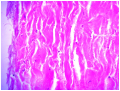
Lyophilized bovine pericardium - was presented as a narrow strip of connective tissue, mainly cell-free substance with no surface epithelium. Somewhere deep in the foundations were examined some fibroblasts with an elongated nucleus and clear cytoplasm. There were no signs of inflammation (Figure 1). When the preparations were stained with hematoxylin and eosin, it was easy to clearly distinguish the fibrous structure of the tissue.

During the immunohistochemical study, the structure of bovine pericardium was represented by loosely arranged bundles of unidirectional fibers. There was a weakly-positive reaction with VEGF and MMP-9, which indicates the slow occurring processes of angiogenesis and fibrosis. The negative reaction to collagen type-IV indicates a high degree of graft extensibility. Also, a negative response was observed with markers CD3 and CD20, suggesting that the LNT has passed multistage process which includes gamma irradiation, which allows to obtain a cell-free substance that contributes to inactivation of the infection.
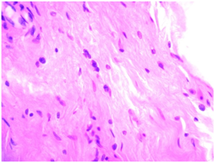
Fascia lata-during histological research was presented as a narrow plate of friable connective tissue stained with eosin evenly, with the presence of a few spindle-shaped fibroblasts with rod-shaped core. No signs of inflammation (Figure 2).
During the iimmunohistochemical study of the reaction with type IV collagen marker was negative, indicating the absence of pathological types of collagen, but for the full picture of the collagen matrix, a reaction to collagen type-I is necessary. There was a weak-positive reaction to CD34 and MMP-9, which indicates the ongoing processes of vascularization and collagenization in transplanted graft. CD3 and CD20 were not detected, indicating that there was no inflammation.
Tunica vaginalis testis - was presented as laminar friable connective tissues that was evenly stained with hematoxylin-eosin during the research, also there were found a few fibroblasts with rounded basophilic nucleus and weakly basophilic cytoplasm. In the thickness of the tissue presented a small focal lymphoid infiltration (Figure 3).

Collagen type IV was detected only in the walls of blood vessels (Figure 4), indicating the process of fibrosis in the thickness of the vascular wall. In the main parts of the thickness of a tissue it was presented rarely (like small islands). Expression of VEGF was also detected focally in the thickness of the drug. The reaction with MMP-9 was moderately-positive, which also indicates the processes of collagenisation. CD34 in the thickness of the tunica vaginalis testis has colored the fibrous fiber. The expression of CD3 and CD20 was weakly-positive, which indicates the presence of inflammatory cells in TVT (Figure 5).
Femoral Vena - smooth muscle cells are found in all three membranes. As a part of the inner and outer membrane were detected longitudinally oriented fascicles of smooth muscle cells, and in the middle membrane-circular fascicles of smooth myocytes separated by loose connective tissue.
During immunohistochemical staining with collagen marker type-IV, its intense expression was determined (+++), which may indicate a poor extensibility of this type of transplant. There was no response from the marker VEGF. CD34 moderately expressed in the capillary endothelium of vasa vasorum. MMP-9 has colored endothelium of larger vessels (Figure 6-8).
The penile deviations, causing erectile dysfunction and unreceptive to conservative therapy, cured today by plaque excision and grafting. One of the most popular grafts today considered to be lyophilized bovine pericardium. This type of graft passes multistage processing in order to minimize antigenic reactions. The process includes gamma irradiation, which allows obtaining a cell-free substance that contributes to the inactivation of an infection. Collagen matrix has the ability to maintain elasticity of original pericardial leaf, it also forms the frame on which the synthesizing new endogenous tissue occurs. The factors that influence on the formation of the frame are the sizes of the transplanted graft and the degree of postoperative inflammation.
Different authors, as grafts to replace the defect of tunica albuginea after excision of fibrous plaques, the following materials have been proposed: fascia lata, the tunica vaginalis testis, femoral Vienna. There are a lot of reports in the literature that contain information of the results of treatment of patients with significant curvature of penis and normal erectile function using the grafts.
LBP Advantages
It passes multistage process, which minimizes the antigenic reaction and inactivates infection; long-term elasticity due to the absence of pathological collagen; there are processes of angiogenesis that indicate the survival rate of the graft.
Disadvantages: High cost.
FLP Advantages
It keeps the elasticity due to the absence of pathological collagen; no inflammation; includes processes of vascularization.
Disadvantages: difficult to predict thickness of the transplanted graft.
TVT Advantages
Thin flap of tissue, availability for sampling the material.
Disadvantages
Inflammation was determined; in vascular walls and in the thickness of the flap fibrosis processes occur; low degree of vascularity and angiogenesis.
VF Advantages
The constant thickness of the transplanted graft.
Disadvantages
The presence of pathological collagen that indicates weak extensibility; possibility of phlebitis development after taking the material; only a narrow strip of tissue can be obtained.
Selecting autoplast for corporoplastic operation must be determined individually taking into account the possible pathological changes in the grafts. Autoplast sizes to close the defect should be selected by taking into account the possible presence of pathological types of collagen.
Postoperative therapy should take into account the processes of vascularization and collagenization of transplanted graft, as well as a possibility of the inflammatory process in autoplast.
None.
The author declares no conflict of interest.

©2017 Andriy, et al. This is an open access article distributed under the terms of the, which permits unrestricted use, distribution, and build upon your work non-commercially.