eISSN: 2471-0016


Case Report Volume 2 Issue 5
1PATOMED Pathology Laboratory, Istanbul, Turkey
1PATOMED Pathology Laboratory, Istanbul, Turkey
Correspondence: Onat Y Akin, PATOMED Pathology Laboratory, Darulaceze cad. No:36/4 Okmeydani- Istanbul- Turkey, Tel +90 212 234 1712, Fax +90 212 234 0854
Received: January 01, 1971 | Published: August 22, 2016
Citation: Akin OY, Genco IS. Giant Ileo-cecal LIPOMA causing intussusception in an adult: a case report and a review of literature. Int Clin Pathol J. 2016;2(5):124–126. DOI: 10.15406/icpjl.2016.02.00056
Intussusceptions in adults are rare conditions with intestinal tumors underlying as the principal cause. Lipomas are an important cause for intussusceptions in adults and they may remain asymptomatic for a long time. The patients usually have non-specific symptoms and the diagnosis relies on imaging methods among which the most useful is CT scan. The treatment consists of surgical intervention to reduce the invagination or to resect the involved segment. In this article we are describing a giant lipoma originating in the distal most ileum, invaginating through the ileocecal valve causing an intussusception of the ileo-cecal segment with total obstruction of the lumen. In our review of the literature it is the largest lipoma of the intestinal tract that has ever been reported.
Keywords: lipoma, giant, intussusceptions, invagination, ileum, cecum, ileo-cecal, ileo-colic, polypoid, tumor
CT, computerized tomography; US, ultrasound
Intussusception is defined as the invagination of a segment of the intestinal tract through and into the lumen of the contiguous portion. It is usually a condition of the pediatric population and the adult form is regarded as a rare condition that constitutes approximately 5% of all intussusceptions and it accounts for 1% of all adult intestinal obstructions.1 Adult intussusceptions are generally the result of benign masses such as various polyps, adenomas and lipomas but malignant lesions such as adenocarcinoma of the bowel are nevertheless may be etiological factors.2 Most studies reported that 60-80% of enteric intussusceptions were benign and 50-65% of colonic intussusceptions were malignant.1
Lipoma of the bowel wall is an infrequent entity and it may very well be a ground for adult type intussusception. They are usually clinically silent and may end up developing intussusceptions when they reach to several centimetres in size. The patients usually complain of abdominal discomfort, colicky pain and have symptoms of acute abdomen. The modalities for diagnoses generally rest on ultrasonography (US) and computerized tomography scan (CT).3
In this article we describe a rare case of a giant lipoma located on the ileo-cecal valve in an adult and ending in a very deforming and obstructive ileo-cecal intussusception. We conclude that our case, lipoma of the intestinal tract measuring 14cm. in the longest axis, is the largest ever reported in the medical literature as compared with the cases described by Namikawa et al.2
A 28 years-old male presented to the general surgery clinic with a one-week history of intermittent abdominal pain, nausea and distention. He had a history of multiple admissions to different hospitals with similar symptoms during this period. On physical examination, diffuse abdominal tenderness, distension and increased bowel sounds on auscultation were found. Abdominal ultrasound was unremarkable. A plain abdominal X-ray revealed distended small bowel loops and air-fluid levels which indicated ileus. (Figure 1) On laboratory blood tests, WBC was 16.3 x 10(9)/L and C-reactive protein was 119mg/L. Obstructive ileus was considered as a diagnosis and surgical intervention was performed to explore the underlying cause for intestinal obstruction. On exploratory laparotomy, a large ileo-cecal intussusception was found with moderate swelling of the ileum. A right hemicolectomy and an ileo-colic anastomosis were performed.
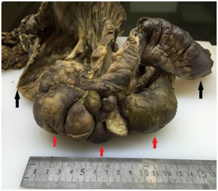
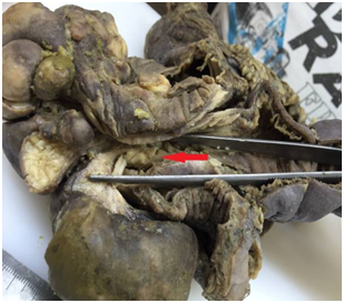
Gross examination of the resected specimen showed an invaginated enteric segment measuring 8x2.5 cm extending from distal ileum to cecum which on cut sections contained a largely pedunculated, yellowish-tan, submucosal polypoid soft tissue tumor measuring 14x7, 5x5 cm in diameter protruding through the ileo-cecal valve and filling and expanding the entire lumen of the cecum. (Figure 2-6). The histology of the polypoid mass showed benign adipocytes proliferating in and confined to the submucosal layer of the ileum and therefore a diagnosis of polypoid ileo-cecal lipoma with intussusception and total obstruction of the cecum was made. The surgical margins of the specimen and a total of 17 lymph nodes were unremarkable. The patient made an uneventful recovery and was discharged six days post-operatively.

Figure 2 Macroscopic features Red arrows indicate the bulge within the ileocecal region. Black arrows indicate ^’’ileum ‘’on the right side and ‘’cecum ‘’on the left side

Figure 3 Macroscopic features Red arrows indicate ‘’the polypoid mass’’. Black arrows indicate ^’’ileum ‘’on the right side and ‘’cecum ‘’on the left side.

Figure 4 Macroscopic features Red arrow indicates the distal ileum containing the mass protruding through the ileocecal valve
Developments of intussusceptions in adults are rare accounting for less than 5% of intussusceptions of all ages and causing 1-5% of all bowel obstructions. In contrast to the much more prevalent pediatric intussusceptions which are idiopathic under etiology in 90% of cases, adult intussusceptions have an organic lesion in 70 to 90% of the cases.4Therefore, the etiology, presentation and management of intussusceptions in adults are different from children. Intussusceptions are classified according to their locations and divided into four categories as enteric, ileo-colic, ileo-cecal and colonic.5 In 44 adult intussusception cases described by Wang et al.,6 enteric and ileo-colic types were the most common ones. Pathological examination of these 44 intussusceptions showed that a tumor occupied 54,5% of the cases, with 27,3%(12/44) being malignant, 25%(11/44) benign, 2,3%(1/44) borderline and 18,1%(8//44) being non-tumoral conditions. 45,4% of these benign tumors which are 11,3 % of all intussusception cases were diagnosed as gastrointestinal lipomas.6
Lipomas of the gastrointestinal tract develop within the mucosa, muscle layer, mesentery, and omentum attached to the intestines. Gastrointestinal lipomas are the second most common benign tumors in the intestine and account for 10% of all benign gastrointestinal tumors and 5% of all gastrointestinal neoplasms. They are most commonly located in the colon (65-75%), in the small bowel (20-25%), and occasionally in the foregut (<5%).3 There are three pathological types of intestinal lipomas according to the location. The submucosal type is the most common, accounting for more than 90% of intestinal lipomas, growing within the submucosal layer, and protruding into the lumen.7,8 The intermuscular type is located within the muscular layer. The subserosal type, which is mostly asymptomatic, grows within the subserosal layer and protrudes out of the gut.9 They are mainly solitary, encapsulated, sessile or pedunculated and their sizes can usually vary from a few millimeters to 5cm or more, median being 3cm.8 Small lipomas are usually asymptomatic and only incidentally detected in colonoscopy or surgery. Lipomas exceeding 2cm diameter usually produce nonspecific symptoms such as abdominal pain, diarrhea or in rare cases acute clinical manifestations may develop due to intussusception or bleeding.5
The clinical presentation of intussusceptions can be very diverse in the adult population with abdominal pain being the most common symptom followed by obstruction and palpable mass. Common physical findings include abdominal distention, hypoactive to absent bowel sounds, abdominal tenderness, heme-positive stools, or an abdominal mass.5,10
Lipomas of the gastrointestinal tract can be diagnosed through conventional endoscopy, capsule endoscopy, barium studies and most importantly CT scan. Typical endoscopic features are a smooth yellowish surface with a pedunculated or sessile base. Other endoscopic characteristics are the “cushion sign” and “naked fat sign.4” Capsule endoscopy and digital balloon endoscopy are newer means for diagnosing lipomas and are particularly helpful in cases involving small bowel lipomas.11 Even with the recent advances of the radiological imaging modalities, intussusceptions are hardly diagnosed preoperatively. Abdominal ultrasound is apt to be masked by gas-filled loops of bowel and hence plain abdominal films are typically the first diagnostic tool since in most cases the obstructive symptoms dominate the clinical picture. Such films usually demonstrate air-fluid levels that suggest an intestinal obstruction and may provide information regarding the site of obstruction.6,12 Even then Barussaud et al.13 reported that the preoperative diagnosis was made in only 52% of patients and Nagorney et al.14reported a low 35%.15 It is also reported that a submucosal lipoma can be diagnosed if a smooth well circumscribed mass of fat density measuring -50 to-100 Hounsfield units is revealed within the lumen of the bowel or intussicipiens.3
Operative intervention is required in all cases of adult intussusceptions and unlike children conservative treatment does not work. The type of procedure depends upon the location of intussusception, pre-operative diagnosis and condition of the intestine at the time of laparotomy.16 Gupta et al.10 reported that small bowel intussusceptions should be reduced before resection if the underlying etiology is suspected to be benign or if the resection required without reduction is deemed to be massive.10
None.
The author declares no conflict of interest.

©2016 Akin, et al. This is an open access article distributed under the terms of the, which permits unrestricted use, distribution, and build upon your work non-commercially.