eISSN: 2469-2778


Research Article Volume 6 Issue 1
1Armed Forces Institute of Pathology, Pakistan
2Armed Forces Institute of Transfusion, Pakistan
Correspondence: Rafia Mahmood, Department of Haematology, Armed Forces Institute of Pathology, CMH Road, Rawalpindi, Pakistan, Tel 923365182270, Fax 92514102783
Received: January 12, 2018 | Published: February 9, 2018
Citation: Mahmood R, Alam M, Altaf C, et al. Phenotypic profile of Rh blood group systems among females of child-bearing age in Pakistan. Hematol Transfus Int J. 2018;6(1):34-37. DOI: 10.15406/htij.2018.06.00148
Objective: To determine the distribution of major Rh antigens and its phenotype in women of child bearing age in Northern Pakistan.
Materials and method: The study was conducted in the Armed Forces Institute of Transfusion (AFIT), Rawalpindi in year 2016.A total of 850 female healthy donors of childbearing age from northern areas of Pakistan between 18 to 45 years were included in the study. Forward and reverse ABO grouping was performed by conventional tube method. Rh phenotyping (for rest of the major antigens of Rh system i.e. C, c, E and e) was done using Bio-Rad gel card.
Results: The phenotypic frequencies of Rh blood groups in the studied population were D-94.94%, C-87.53%, E-21.41%, c-61.18% and e-98.59%. Thus ‘e’ was the most common and E was the least common of all the Rh types. Phenotypically DCCee group was the most common phenotype.
Conclusion: Determination of population based frequency data of Rh antigens and phenotypes can play a major role in predicting and managing haemolytic disease of the fetus and newborn (HDFN) and preventing alloimmunization and avoiding adverse events in multi-transfusion cases.
Keywords: Rh blood group, Rh phenotypes, Pakistan
The Rh blood group system discovered in 1939 is only second in importance to the ABO system in terms of transfusion.1 It is a highly complex blood group system with more than fifty different antigens.2 However, most clinical transfusion issues are associated with the five principal antigens D, C, c, E and e. Rh antigens are non-glycosylated proteins in the RBC membrane.3 Two closely linked genes on chromosome 1 control the expression of Rh; one gene RhD codes for the presence of RhD, and a second gene codes for the expression of CcEe antigens.4 The Rh antigens are thought to play a role in maintaining the integrity of the RBC membrane. They may also be involved in transport of ammonium across the RBC membrane.5
Red blood cell stimulation through transfusion or pregnancy leads to antibody production.4 Most Rh antibodies are IgG immunoglobulin and can cross the placenta.6 Rh antibodies have been implicated as a clinically significant cause of hemolytic disease of the fetus and newborn (HDFN) and hemolytic transfusion reactions (HTR), usually delayed transfusion reactions.7 Rh antibodies do not bind complement and Rh mediated hemolytic transfusion reactions usually result in extra vascular hemolysis.8 HDFN occurs due to the maternal IgG antibody that crosses the placenta. This antibody binds to the fetal antigen-positive RBCs, thus leading to their hemolysis.9 Of all the RBC antigens, D is the most immunogenic and responsible for the most severe cases of HDFN.10 The introduction of RhIG (Rh immune globulin) has reduced the incidence of Rh HDFN. Anti-c is the second most common cause followed by anti-E.11
Blood group distributions are different and distinctive for population groups, and can show noticeable differences in geographic prevalence around the world.12 The present study was designed with an aim to see the prevalence and distribution of the Rh antigens in our part of the country as so far there is lack of data on the frequency of Rh phenotypes in our region. Knowledge of the distribution of Rh antigens in our population can be useful for blood banks to maintain their inventory, to find antigen negative blood for alloimmunized patients, to calculate the number of blood units to be cross-matched to find a compatible unit and for the preparation of indigenous cell panels. It is also important as it helps to predict HDFN. This study provides a database to blood banks and transfusion centers for searching antigen negative blood for alloimmunized patients and for the preparation of indigenous cell panels.13
Subjects
A total of 850 healthy females from northern areas of Pakistan were randomly selected for red cell Rh phenotyping. They belonged to different ethnic groups including Pathans, Punjabis, Sindhi, Balochis and people from Gilgit Baltistan. Patients were between the ages of 18-45years. Blood samples were collected from females who came for routine blood grouping, females coming to gynecology and obstetrics outpatient department and indoor patients admitted in gynecology and obstetrics ward. Females with no previous history of any transfusion were included only. All subjects were elaborately apprised about the study and written informed consent was obtained.
Blood grouping and Rh phenotyping
A 5 ml sample of blood was drawn from antecubital vein and 2ml transferred immediately to a tube containing EDTA and 3ml collected in plain tube. Forward and reverse ABO grouping was performed by conventional tube method. Rh D typing was done by tube method using monoclonal/polyclonal anti-D. Rh phenotyping (for rest of the major antigens of Rh system i.e. C, c, E and e) was done using Bio-Rad gel card according to manufacturer instructions.
Data analysis
All the collected data were entered in statistical package for social sciences (SPSS) version 20. The analyzed variables included D, C, c, E and e antigen and allele frequencies were calculated from these. Phenotype were observed, noted and expressed as percentages and most probable genotypes were determined from gene frequency estimates. Data was entered and analyzed using SPSS version 20.
Ethical approval
This study was approved by the Ethical Review Committee of Armed Forces Institute of Transfusion, Rawalpindi. Informed written consent was taken from the patients.
A total of 850 patients were included in this study. Mean age of the patients was 28.16±8.30 years (ranging from 18-45 years). ABO grouping in this study showed that the most common blood group was B seen in 285 (33.53%), followed closely by blood group O in 252 (29.65%) of the sample population. Blood group A was seen in 230 (27.06%) of the females with blood group AB in 83 (9.76%) being the least common. In the Rh blood group, 807 females representing 94.94% of the sample population were found to be RhD positive while 43 (5.06%) of the females were RhD negative. Weak D was not detected in any of the females.
Of the five antigens that were phenotyped, the e antigen was found to have the highest frequency (98.59%), followed by D (94.94%) and C (87.53%) antigens as shown in Table 1. The lowest prevalence was observed for the E antigen (21.41%). The most common phenotype was DCCee (38.82%), followed by DCcee (32.71) and DCcEe (14.24%). The least common phenotypes were dccee (2.71%), Dccee (2.6%), dCcee (1.76%), DccEE (1.4%), dccEe (0.6%). No case of phenotype dCcEe and dCCEE was detected (Table 2).
Red Cell Antigen |
No of Persons (N=850) |
% |
D |
807 |
94.94 |
d |
43 |
5.06 |
C |
744 |
87.53 |
c |
520 |
61.18 |
E |
182 |
21.41 |
e |
838 |
98.59 |
Table 1 Frequency of red cell Rh antigens in the study population
Results with Anti-Sera |
Phenotype |
Frequency |
|||||
D |
C |
E |
c |
e |
N=850 |
% |
|
+ |
+ |
- |
- |
+ |
DCe |
330 |
38.82 |
+ |
+ |
- |
+ |
+ |
DCce |
278 |
32.71 |
+ |
+ |
+ |
+ |
+ |
DCcEe |
121 |
14.24 |
+ |
- |
+ |
+ |
- |
DcE |
12 |
1.4 |
+ |
- |
- |
+ |
+ |
Dce |
22 |
2.6 |
+ |
- |
+ |
+ |
+ |
DcEe |
44 |
5.18 |
- |
- |
- |
+ |
+ |
Ce |
23 |
2.71 |
- |
+ |
- |
+ |
+ |
Cce |
15 |
1.76 |
- |
- |
+ |
+ |
- |
cE |
5 |
0.6 |
Table 2 Reaction pattern with antisera, phenotype and probable genotype frequencies in the study population
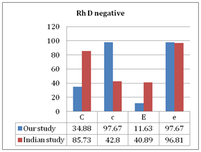
We also compared the frequency of these Rh antigens (C,c,E,e) in RhD positive and RhD negative females in the study population. In RhD positive females, antigen e (98.51%) followed by C (90.33%) was most frequent while in RhD negative females, the most frequent antigens were e (97.67%) and c (97.67%).
Among the blood group systems, after the ABO, Rh is the second most important blood group system.11 ABO antibodies are typically found in individuals who lack the corresponding antigen but in the Rh system, it is only after exposure to foreign red blood cell antigens that Rh antibodies are formed.2 The exposure to red cell antigens may be through transfusions or pregnancies.12 Once alloantibodies against foreign antigens are made, they can produce significant hemolytic disease of the fetus and newborn and hemolytic transfusion reactions.6 Karim et al.9 in a study conducted in Pakistan has reported the risk of sensitization with D antigen in D negative women to be as high as 2.2%.9
There are very few studies from Pakistan on Rh phenotype frequencies. Armed Forces Institute of Transfusion is a big institute in north of the country. It caters to a large number of people from all over the country from very different ethnic backgrounds, including patients from tribal areas and Peshawar which are Pathan in origin and patients from Gilgit, Baltistan and Abbottabad and from north of Punjab. This study helps us to determine the Rh antigen prevalence in our population, thus providing a database to blood banks and transfusion centers for predicting the alloantibodies formed in patients receiving transfusions, predicting HDFN, searching antigen negative blood for alloimmunized patients and for the preparation of in-house panel cells.
ABO grouping in this study showed that B(33.53%) was the most common blood group, followed by O(29.65%) while AB(9.76%) was the least common. These findings are in accordance with an Indian study by Garg et al.14 that has reported B to be the most common blood group in the Indian population and AB to be the least common. However, blood group O has been reported to be the most common in population from southern Pakistan in a local study by Karim et al.15
Our result of 94.94% RhD positive persons is in agreement with a study by Sarkar et al.16 (92.25%) and Das et al.17 (94.53%). However, Fisher et al.18 has reported a frequency of 83-85% in Europeans and Americans and a frequency of 99% in the Chinese population. Of the Rh antigens, the e antigen (98.59%) had the highest frequency, followed by C antigen (87.53%). Our results match with those of another local study by Karim et al.15 who has reported e antigen-99% and C antigen-87%. Frequency of e antigen-97.9% and C antigen-75.9% has been reported in the Iranian population by Keramati et al.19 Table 3 shows the frequency of the Rh antigens as reported in different studies.
Antigen |
Our Study Pakistan % |
Karim et al.15 Pakistan |
Keramati et al.19 Iran |
Bogui et al.20 Africa |
D |
94.94 |
97 |
90.2 |
92.93 |
C |
87.53 |
87 |
75.9 |
21.97 |
c |
61.18 |
57 |
73.9 |
99.85 |
E |
21.41 |
19 |
29.5 |
13.82 |
e |
98.59 |
99 |
97.9 |
99.85 |
Table 3 Comparison of frequency of Rh antigens in different populations in the world
The antigen frequencies of other Rh antigens (C, c, E, e) in D positive individuals and their frequencies in Rh D negative individuals are compared with those of the Indian population16 in Figure 1 & Figure 2 respectively. The most common Rh phenotype in our population is DCCee (38.82%), followed by Dccee (32.71%). These findings are consistent with those reported by Sarkar et al.16 He has reported the frequency of DCCee as 35.2%, the most frequent in the Indian population and Dccee as 30.7%, second most frequent in their population. However, the frequency of DCCee is 17.6% in Whites and 2.9% in Africans, while the frequency of Dccee is 31.1% in Whites and 8.8% in the African population11–13 as shown in Table 4.
Phenotypes |
Our Study Pakistan % |
Karim et al.15 Pakistan |
Sarkar et al.16 India |
African Blacks 11-13% |
Whites 11-13% |
DCe |
38.82 |
41 |
35.2 |
2.9 |
17.6 |
Dce |
32.71 |
34 |
30.7 |
8.8 |
31.1 |
DCcEe |
14.24 |
10 |
8.1 |
3.7 |
11.8 |
DcE |
1.4 |
1 |
0.7 |
1.3 |
2 |
Dce |
2.6 |
3 |
2.2 |
22.9 |
3 |
DcEe |
5.18 |
8 |
5.9 |
5.7 |
10.4 |
Ce |
2.71 |
1 |
0.3 |
7 |
15 |
Cce |
1.76 |
1 |
2.5 |
1 |
1 |
cE |
0.6 |
1 |
Rare |
Rare |
1 |
Table 4 Comparison of Rh phenotype frequencies in different populations in the world

Figure 2 The comparison of frequency of Rh antigens in Rh D negative individuals in our study and an Indian study.16
This study has documented Rh antigen frequency in population of Northern Pakistan. We have compared our data with Iranians, Africans and Indian population and found that the frequencies in our population resemble those in Indians and Iranians. The data are useful not only for creating a donor data base and finding antigen negative compatible blood for alloimmunized patients but also for the preparation of indigenous panel cells. We have performed only serological investigations and as the gene frequency of Rh antigens is not clearly known in our population, so further studies including molecular investigations are required on this subject using large sample size to establish the genetic make-up of Rh system in our population.
We are grateful for the technical support provided by Muhammad Yaqoob.
No potential conflicts of interest relevant to this article were reported.

©2018 Mahmood, et al. This is an open access article distributed under the terms of the, which permits unrestricted use, distribution, and build upon your work non-commercially.