eISSN: 2469-2778


Case Report Volume 4 Issue 5
1Department of Virology & Immunology, Haffkine Institute, India
2Department of Microbiology, Sir JJ Hospital, India
Correspondence: Habip Gedik, Department of Infectious Diseases and Clinical Microbiology, Ministry of Health Bakırköy Sadi Konuk Training and Research Hospital, Turkey, Tel 90 212 314 55 55
Received: April 10, 2017 | Published: May 30, 2017
Citation: Yokus O, Gedik H, Sahin EC. Practical staining method of well-unstained haematological specimens. Hematol Transfus Int J. 2017;4(5):136-137. DOI: 10.15406/htij.2017.04.00098
In hematology, May-Grunwald-Giemsa (MGG) staining is used to evaluate bone marrow aspirate as well as blood smear. This is an easy method in order to avoid the damage of the cells; good qualities of the stain and its directives, as well as the duration of the procedure are very important. The smears need to be dried quickly after the procedure. For a successful smear needs to be thin and while drying it up, hot blowing can be used. In case bone aspirate or blood smear are not well stained by MGG, the aspiration has to be re-executed or the biopsy results should be awaited. That procedure takes approximately 15days that is needed to get the biopsy result and a long time that leads to material and motivational loss. In our clinic, we use a practical stain method for pre-diagnosis in such cases. Two cases are presented in this study inform you about that method that yielded good results on a first trial.
Keywords: may-grunwald-giemsa, bone marrow aspirate, pale staining, lamella, biopsy
Case 1
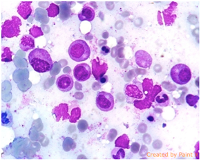
28 years old male patient diagnosed with Acute Myeloid Leukemia had 3+7 chemotherapy. On the 28th day of the treatment after the peripheral blood smear results improved; bone marrow aspirate was pale, although all the cells were stained (Figure 1). The second stain was performed and the results were perfect. Less than 5% of blasts were seen supporting the idea of remission (Figure 2).

Figure 1 Control of AML case after remission induction therapy. Bone marrow sample was bad stained and cells do not look good.

Figure 2 After the second staining of the same sample of AML case, the cells of Figure 3 look better.
Case 2
We were asked for consultation of 38 years old pancytopenia patient from department of internal medicine. On the first try of the staining, we could not get the result due to pale staining (Figure 3). The second staining was performed and results were satisfied (Figure 4).
Although there is a little difference among medical centers, the summary of the techniques of MGG staining is like that: Bone marrow aspirate or blood smear which is dried in the ambient temperature is covered pouring May-Grunwald (5-7drops) and then it is waiting two minutes. Then it is poured away (without washed by tap water) and prepared Giemsa staining (5-7drops) with distilled water is poured onto lamella waiting for 7minutes. Then lamella is washed with tap water that is quickly dried and examined under the microscope. With this technique, if the lamella is not stained well, or the sample is pale, our experience is that those samples could be stained well executing shorter staining time with less amount of dye. We pour over the samples with May-Grunwald for 1.5min and then pour cover them with Giemsa for 4min. After that, we wash the lamella in the same way under tape water, dry up and then examine. As seen in four pictures, the samples that could not be examined are well stained for examination.
As a result, when an insufficiently stained sample is dyed again with the same stain procedure in shorter period, it is morphologically evaluated better. This method prevents a great waste of time and suffering of patient with a re-biopsy.1 In practical hematology applications, this procedure will be useful and provide exact results. As we want to share this practical method with our colleagues, although there may be some with different experiences.
None.
The author declares no conflict of interest.

©2017 Yokus, et al. This is an open access article distributed under the terms of the, which permits unrestricted use, distribution, and build upon your work non-commercially.