eISSN: 2576-4497


Research Article Volume 5 Issue 1
1School of Medicine, University Islam Bandung, Indonesia
2General Surgeon, RSUD Sultan Syarif Mohamad Alkadrie, Indonesia
Correspondence: Hendro Sudjono Yuwono, School of Medicine, University Islam Bandung, Bandung 40116, Indonesia , Tel +62811206443
Received: June 07, 2022 | Published: July 25, 2022
Citation: Yuwono HS, Nugroho BE. Coffee prevents damaged of wound bed cells. Hos Pal Med Int Jnl. 2022;5(1):8-11. DOI: 10.15406/hpmij.2022.05.00199
Objectives: The superficial wound cells adhered to the dressing during replacement and may delay wound healing. It is different from using the coffee ground as a wound dressing. The coffee has an impressive antioxidant, anti-inflammatory, and antibacterial power. Some of it stayed on the wound to protect the proliferating cells. Are there any wound cells shed when dressing change using coffee ground?
Methods: We conducted a prospective observational study in Wistar rats as an experimental randomized control trial using 27 rats of 250 grams divided into three groups. The wound size was 2cmx2cm, deep 2 mm at the back skin, and closed using gauze-saline (Group-1), foam dressing (Group-2), and coffee powder (Group-3). On the 7th day, the dressing was fixed using Liqui-PREP solution to make histologic slides to counting the epithelial cells using a microscope with 200x magnification. The wound biopsied for histologic assessment. Scoring of attached cells in wound dressing: < 10/HPF= score 3, 10-20/HPF= score 2, >20/HPF = score 1. The healing of collagen tissue density 0 to 50% = score 1, 50-75% =score 2, 75-100%=score 3. The healing of epithelialization density 0 to 50% = score 1, 50-75%=score 2, 75-100%=score 3. The healing of new vascular density, none = score 1, 1-2 new vascularization = score 2, more than 2 of new vascularization = score 3.
Results: The scoring of attached epithelium in Group-1: 1.55±726, Group-2: 2.55±0.527, Group-3: 3 (p=0.0001). The coffee powder group was the significant least attached cells.
Wound healing rate, Group-1: 5.77±0.44; Group-2: 6.55±0.88; Group-3: 7.22±1.20 (p=0.012). Collagenization in Group-1: 1,33±0,50, Group-2: 1.78±0.67, Group-3: 2.67±0.50 (p=0.001). Epithelialization in Group-1: 1,77±0,66, Group-2: 1.88±0.60, Group-3: 2.11±0.60 (p=0.004). Vascularization, in Group-1: 2.33±0,50; Group-2: 2.10±0.60; Group-3: 2.78±0.44 (p=0.041).
Conclusions: The coffee powder has minimum detached epithelial cells.
Keywords: coffee powder, foam, gauze-saline, wound healing, attached epithelium
The village people in the coffee estate have used handcraft coffee for a long time to treat injuries. The deep-rooted experience recognizes its validity in wound healing. Our task is to prove the performance of the coffee accurately. One of the evidentiary efforts is the research below. At present, the management of wounds might cause discomfort due to repeated debridement and frequent dressing replacement, costly and atrocious. The previous experiments showed that coffee powder could faster healing than gauze, topical antibiotics (Bacitracin-Neomycin powder, Gentamycin sulfate), Ag-sulfadiazine, foam pad, and hydrocolloid.1
The study was approved by the institution’s ethical clearance committee decree, including animal welfare regulations of the institution according to the guidelines of the Helsinki declaration.
We conducted a prospective observational study in Wistar rats as an experimental randomized control trial using 27 rats of 250gram divided into three groups.
Group-1: gauze of 3cmx3cm soaked in 5 ml 0.9%NaCl.
Group-2: a foam pad dressing (Wundres Nâ, Biopol, South Korea) measured 3 cm x 3 cm.
Group-3: 5gram of coffee powder for every wound and closed with dry sterile gauze and adhesive tape to hold the powder from spilling.
Animal study
Inclusion criteria: male Wistar rat, three-month-old, 250-gram, has been quarantined for one week.
Exclusion criteria: unhealthy rat or died, less than 250 grams during the experiment.
All animal study held in the Pharmacology laboratory, School of Medicine, Universitas Padjadjaran, Indonesia.
Three groups, nine rats per group selected using simple random sampling. Every two rats were put in one cage under standardized room temperature, humidity, and light conditions every 12 hours cycle with free access to pellets and water.
Ketamine HCl (Ivanesâ, PT.Ikapharmindo pharmaceutical industry) 80mg/kg body weight intraperitoneal injection, hair shaving, and made of the wound size 2 cm x 2 cm with the depth of 2 mm on the back-side. All dressings were covered with sterile gauze and fixed with adhesive plaster tape (Johnson & Johnson, USA). The wound dressing changed on the 7th day.
Histological examination
On the7th day, all dressings were removed and fixed into the Liqui-PREP solution (LGM International Inc., USA), as shown in Figure 1. The gauze attached to the wound is fixed in the Liqui-PREP liquid; then the liquid is shaken using a vortex for 10 seconds, then 10 minutes centrifuge of 2000 rpm. The sediment was taken to be made slides with the Papanicolaou staining for the assessment using a light microscope. A pathologist counted the cells using a microscope (200 x-magnification). Epithelial cells attached to wound dressing (Table 1): less than 10 epithelial cells/HPF = score 3, 10-20 epithelial cells/HPF= score 2, more than 20 epithelial cells/HPF = score 1 (HPF=high-power field).

Figure 1 Three different dressings were applied closed with sterile gauze and fixed with the adhesive plaster. The wounds did not wash during dressing replacement, and the coffee powder was also not cleaned at the wound surface. The thin layer of coffee remains at the superficial during dressing replacement. It will prevent any trauma to the wound surface and keep the growing cells undisturbed. The dressing was fixated in Liqui-PREP and prepared to make slides for microscopic examination.
Attached cells / HPF |
score |
<10 cells |
3 |
10-20 cells |
2 |
>20 cells |
1 |
Table 1 Attached cells and score
The wound biopsy examination was done applying the score shown in Table 2. The biopsy specimen was put into 10% formalin solution for 18-24 hours, then stained using the Hematoxylin-Eosin stain. Collagen density 0-50% normal average score 1, collagen density 50-75% normal average score 2, collagen density 75-100% normal average score 3. Epithelial growth 0-50% score 1, epithelial growth 50-75% score 2, epithelial growth 75-100% score 3. No new blood vessel has grown score 1; one or two new blood vessels appear score 2; more than two new blood vessels appear score 3. A senior pathologist carried out the histologic assessment in the randomized, double-blind method (the pathologist did not know the slide belonged to which group).
Degree |
Histologic examination of wound biopsy |
Score |
1 |
Collagen density 0-50% normal |
1 |
2 |
Collagen density 50-75% normal |
2 |
3 |
Collagen density 75-100% normal |
3 |
1 |
Epithelialization 0-50% |
1 |
2 |
Epithelialization 50-75% |
2 |
3 |
Epithelialization (75-100%) |
3 |
1 |
No new blood vessel |
1 |
2 |
1-2 new blood vessels |
2 |
3 |
More than 2 new blood vessels |
3 |
Table 2 Histologic examination and score
Biopsy of the wound
The wound was biopsied and fixated in formalin, and processed into slides. A senior pathologist applied light microscopic examination to assess the growth of epithelium, collagen, and new vessel.
The coffee powder:
We bought the coffee (Coffea arabica) from a coffee shop at Banceuy street no.51, Bandung, Indonesia.
We bought the gauze and physiologic saline from PT. Kimia Farma pharmaceutical industry, Indonesia.
Statistical analysis
The statistical data was analyzed using SPSS 23.0 for Windows. For non-categorical data or quantitative data scales used One Way Anova in normally distributed data, instead used Kruskal-Wallis statistic on abnormal data. Categorical data of p-value are calculated based on the Chi-Square test with alternative Kolmogorov-Smirnov test and Exact Fisher test if the requirements of Chi-Square statistical test are not met. The p-value <0.05 considered significant.
Cells attached to wound cover
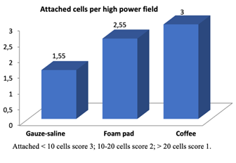
Table 1 & 2 mention the score of attached cells and the degree of healing. Figure 1–3 show the new cells on the wound surface released, adhered to the wound dressing, and the degree of wound healing. The number of cells attached in the coffee group, is less than in the other group, and better recovery. At the time of replacement deliberately maintained a layer of coffee powder was to the wound surface to protect the growing cells. Table 3 & Figure 2 show the attached epithelium in Group-1 was 1.55±0.726, in Group-2 was 2.55±0.527 and in Group-3 was 3 (p=0.0001), with a score 3 as the maximum score.
Variable |
Epithelial cells adhered per HPF |
P |
||
Mean±Std |
Median |
Range |
||
Gauze-saline |
1.55±0.726 |
1.00 |
1.00-3.00 |
|
Foam pad |
2.55±0.527 |
3.00 |
2.00-3.00 |
0.0001 |
Coffee |
3 |
- |
- |
|
Table 3 Epithelial cells adhered to wound dressing per HPF

Figure 2 The attached epithelial cells per high power field. The coffee group was the least number of cells attached to the wound dressing.

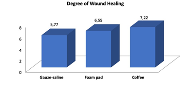
Figure 3 The degree of wound healing. The coffee group was the higher healing degree compared to other dressing.
Wound healing
In Table 5, and Figure 3, the rate of wound healing in Group-1 was 5.77±0.44; Group-2 was 6.55±0.88, and Group-3 was 7.22±1.20 (p=0.012). Table 4 showed the difference between the two groups, and the coffee group was the less adhered cell. Degree collagenization (Table 6, Figure 4) in Group-1 was 1,33±0,50, Group-2 was 1.78±0.67, and Group-3 was 2.67±0.50 (p=0.001).
Variable |
Epithelial cells adhered to wound per HPF |
P |
||
>20 cells |
10-20 cells |
<10 cells |
||
Gauze-saline |
5(55.6%) |
3(33.3%) |
1(11.1%) |
0.124 |
Foam pad |
0(0.0%) |
4(44.4%) |
5(55.6%) |
|
Gauze-saline |
5(55.6%) |
3(33.3%) |
1(11.1%) |
0.002 |
Coffee |
0(0.0%) |
0(0.0%) |
9(100.0%) |
|
Foam pad |
0(0.0%) |
4(44.4%) |
5(55.6%) |
0.082 |
Coffee |
0(0.0%) |
0(0.0%) |
9(100.0%) |
|
Table 4 The amount of epithelium attached to the wound dressing in two groups
Variable |
Wound healing rate |
P |
||
|
Mean±Std |
Median |
Range (min-max) |
|
Gauze-saline |
5.77±0.440 |
6.00 |
5.00-6.00 |
|
Foam pad |
6.55±0.881 |
7.00 |
5.00-8.00 |
0.012** |
Coffee |
7.22±1.201 |
7.00 |
5.00-9.00 |
|
Table 5 Comparison of the Degree of Wound Healing in the Gauze-saline, Foam pad, and Arabica Coffee Powder groups
For numerical data, the p-value is tested by using the One Way Anova test if the data is normally distributed with the alternative of the Kruskal Wallis test if the data is not normally distributed. The significance value is based on the p-value <0.05. The * sign indicates the value of p<0.05, which means that it is statistically significant or significant.
Variable |
Collagenization |
P |
||
Mean±Std |
Median |
Range(min-max) |
||
Gauze-saline |
1.33±0.500 |
1.00 |
1.00-2.00 |
|
Foam pad |
1.88±0.600 |
2.00 |
1.00-3.00 |
0.005 |
Coffee |
2.33±0.500 |
2.00 |
2.00-3.00 |
|
Table 6 The collagenization
Degree epithelialization (Table 7, Figure 5) in Group-1 and Group-2 were 1.89±0.60, Group-3 was 2.78±0.44 (p=0.004). Degree vascularization (Table 8, Figure 6) showed Group-1 was 2.33±0,50, in Group-2 was 2.10±0.60, Group-3 was 2.78±0.44 (p=0.041).
Variable |
Degree epithelization |
P |
||
Mean±Std |
Median |
Range(min-max) |
||
Gauze-saline |
1.77±0.666 |
2.00 |
1.00-3.00 |
|
Foam pad |
1.88±0.600 |
2.00 |
1.00-3.00 |
0.5 |
Coffee |
2.11±0.600 |
2.00 |
1.00-3.00 |
Table 7 The degree of epithelization
Variable |
Degree of new vascularization |
P |
||
Mean±Std |
Median |
Range(min-max) |
||
Gauze-saline |
2.66±0.500 |
3.00 |
2.00-3.00 |
|
Foam pad |
2.77±0.440 |
3.00 |
2.00-3.00 |
0.831 |
Coffee |
2.77±0.440 |
3.00 |
2.00-3.00 |
|
Table 8 The degree of new vascularization
The caffeinated coffee powder is a topical wound cover that can accelerate the growth of new cells in the therapeutic performance. The community experiences and the author’s 15 years of clinical investigation in using coffee to treat wounds have shown its success.1 It’s mainly for the high ability of anti-bacterial, antioxidant, anti-inflammatory, and spark skin microcirculation.1,2 The coffee powder can manage through all stages of the therapeutic injury process and shorted the inflammation, collagen proliferation, and epithelialization to quicker healing.1 Coffee powder is almost available everywhere and is straightforward to apply.1
The superficial wound cells made adhesion and clung to the dressing. Thus, frequent dressing replacement may be traumatic to the wound bed, especially by the adhered gauze, which creates a healing delay. Coffee powder treated the wound, and the superficial cells will not cling to the dressing because a thin layer of coffee powder deliberately remained on the wound surface during and after replacement.
Winata et al.3 reported that coffee creates a decreased MMP-1 level in wounds infected by Staphylococcus aureus ATCC 25923. The coffee was a robust anti-inflammatory, and conceived the fastest wound healing compared with hydrocolloids (statistically not significant) or gauze (statistically significant) in the rat.3
Repeated unnecessary debridement should not be done because harmful and traumatic to the wound tissue except for careful removing dead tissue. The repeated washing with saline or distilled water will elevate the pH of the wound tissue and cause bacterial contamination, which allows a new inflammation.4,5 The bacteria could enter the wound from the hair follicles of the neighboring healthy skin while wetting the damage.5
The ideal topical wound dressing should have significant abilities, particularly antibacterial, anti-inflammatory, antioxidant, economical, and activate skin microcirculation.1 That the coffee powder has. And ultimately, it can cure with a straightforward and efficient procedure.1
During every dressing replacement, the superficial cells clung to the dressing. Thus, frequent dressing replacement of an adherent dressing causes trauma to the wound surface’s cells, primarily by sticky gauze, and as a consequence, there is slower healing.
Repeated debridement should perform with care when necessary to avoid the death of new growing cells.
Figure 3 shows that the loss of epithelial cells by lifting the dressing will always be a release of the cells except using coffee powder.3,6 The more frequent replacement, the more cell injury occurs.5 Any wound dressing allows damage to cells due to its adhesive properties. Although the dressing stickiness is relatively present, the cells will be loose due to frequent replacement. Every day replacement causes the shed cells every day as well. Cell damage is exacerbated by washing and rubbing the wound. Sticky wound dressings (gauze, etc.) damage cells. However, wound healing occurs. Healing occurs because there is a repopulation of existing and healthy cells. But it will take more time. A layer of coffee guards the wound surface against any trauma, resulting in speed healing.7 The action of coffee is significantly helpful compared to other wound dressings.7 Maintenance of cells that grow on the surface of a wound is essential for healing performance.
The coffee powder has minimum detached wound cells and the fastest healing compared to gauze and foam dressing. The utilization of non-adherent dressing anticipates injury to new growth of epithelial cells. A thin layer of residual coffee powder left behind on the wound surface will prevent wound dressing adhesion, protect the cell’s growth, non-traumatic, and accelerate proliferation. Using coffee with antioxidant, anti-inflammatory, and antibacterial properties is effective in wound healing.
None.
The authors declared no conflicts of interest.

©2022 Yuwono, et al. This is an open access article distributed under the terms of the, which permits unrestricted use, distribution, and build upon your work non-commercially.