eISSN: 2469-2794


Research Article Volume 2 Issue 4
1Dental Care and Research Center, India
2Department of Oral Pathology, Sri Siddhartha Dental College, India
3Sri Siddhartha Dental College, India
Correspondence: Charan Gowda BK, Department of Oral Pathology, Sri Siddhartha Dental College Tumkur, Karnataka State, India -560004
Received: June 20, 2016 | Published: July 7, 2016
Citation: Mohan CV, Charan GBK, Denish SH, et al. Postmortem histological changes of oral mucosal tissue in hypoxic and histotoxic death to differentiate cause and manner of death. Forensic Res Criminol Int J. 2016;2(4):149-151. DOI: 10.15406/frcij.2016.02.00062
Cell is the basic structural and functional unit of all living organisms. In biological aspects humans though consider to be similar to other living organisms, they are unique as humans can think, imagine, and create so on. It is necessary to know the accurate cause of the death for solving many of the legal and administrative problems. In many cases cause of the death is determined by medical investigator.
Aims & objective: To study microscopic changes in oral mucosal tissue from cases of hypoxic and histotoxic death.
Materials & method: Fifteen oral mucosal tissue samples each form poisonous and hanging death were collected during autopsy. Tissues were routinely fixed, processed and stained using Haematoxylin and Eosin stain. Sections were studied and nuclear and cytoplasmic changes of epithelial cells were observed.
Results: Definitive changes like prominent cytoplasmic vacuolation, in case of hanging and nuclear morphological changes in cases of poison death were observed.
Conclusion: Biochemical mechanism behind cell death varies with cause of death, which in turn can be observed as morphological changes.
Keywords: homeostasis, hypoxic hypoxia, anemic hypoxia, stagnant hypoxia, histotoxic hypoxia
Cell is the structural and functional unit of life. It is composed of nucleus and cytoplasm. In living individuals cell will communicate constantly with the signals it receives from surrounding environment. A normal cell receives nutrients and converts into energy to perform its function and maintain a steady state called homeostasis.1,2 Stress and stimuli bring about certain morphologic adaptations within the cell. If the cell fails to adapt to any stress and stimuli it can result in cell death. various morphological changes like enlargement of cell, disruption of cell membrane and nuclear Pyknosis, karyorrhexis, karyolysis may occur which depend on the type of injury, its duration, and its severity.3 Cell death can occur due to various reasons more common being - hypoxia, ischemia, toxins, infection etc. When the changes occur in cell due to injury and persist for a longer period, cell death occurs.4-8 Within Merriam Webster dictionary they define death as “the irreversible cessation of all vital functions especially as indicated by permanent stoppage of the heart, respiration, and brain activity”.9 Death is inevitable and occurs in every living organism including humans. After death, a sequence of changes naturally occurs in the human body which can be made out grossly. These gross changes occur because of changes at the cellular level.
Before cell injury/cell death, cell tries to adapt to stimuli. When injury is severe enough or cell fails to adapt to the injury it reaches to a “point of no return” or cell death.3
Cause of death is defined as, the mechanism leading to alterations in physiology and biochemistry of individual cell resulting in death. Manner of death is a sequence that produces fatality without which the end result would have not occurred. Cause of death can be established based on the histological changes of cell, whereas manner of death can be established by visual appearance. It is very much needed to know cause of death in many legal and administrative purposes.10,11 Causes of hypoxia can be divided into four main categories: Hypoxic (anoxic), Stagnant (Shock, Ischemia), Histotoxic and Anemic (reduced oxyhaemoglobin content).3 The manner in which death occurs following which cell injury or cell death results can be grouped as Oxygen deprivation, physical agents like mechanical trauma, chemical agents/ toxins, infectious agents and nutritional imbalances etc.3 In modern days, society is facing a challenge in solving many crime one such is to differentiate the histotoxic death and respiratory failure death. Considering all the above factors mentioned about cell death we attempted to differentiate between hypoxic and histotoxic death by observing histological changes of oral mucosa. Proper differentiation of cause and manner of death will help legal as well as administrative agency to solve many problems.
Cross sectional study using convenience sampling was carried for period of one year from 01/05/14 -31/04/15. Institutional ethical clearance for the study was obtained. Tissue samples were collected from the persons who were brought dead to the hospital for autopsy due to poisoning or hanging. Samples were obtained within 10th hour following death during autopsy. Total of thirty deceased individual were selected for study. Among which fifteen oral mucosal samples was from poisonous death and fifteen more was hanging death were included in the study and other causes were not considered. Tissue samples of size 5mm x 5mm x 5mm were obtained from middle third of tongue from dead persons during autopsy procedure performed at Department of Forensic Medicine. Informed consent was obtained from the legal representative of the dead person. Data regarding the deceased individual like age, gender, estimated time of death was recorded. Autopsy tissue samples were collected within 6-12hrs after death. Tissue samples were fixed in 10% formalin. Tissue was routinely processed and stained with Haematoxylin and Eosin stain.12 The stained sections were observed microscopically and examined for changes in the shape of cell, epithelial cell membrane, shape of nucleus and nuclear membrane at Department of Oral & Maxillofacial Pathology.
Of the fifteen each case studied in both the groups all the cases showed the epithelial and nuclear changes. Nuclear, cytoplasmic vacuolation was also noted.
In hypoxic death
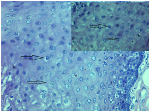
Generalized cytoplasmic vacuolation along the basal and supra basal layers were evident. It was also observed that spinous cells showed edematous changes. Cytoplasmic membrane and nuclear membrane were discernible.
In histotoxic death
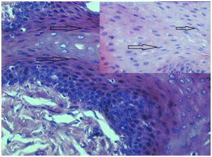
Though cytoplasmic vacuolation was noted it was not as prominent as in hypoxic death. Cytoplasmic membrane and nuclear membrane were not discernible. Prominently noted feature among the poisonous death was presence of arc shaped nucleus.
Cell being a basic living unit of life, takes up oxygen which reacts with carbohydrate, fat, and protein to release energy required for its function. Different chemical reactions occur within the human cell in order to maintain body function. Cells perform their function till they receive proper concentrations of oxygen, glucose, different ions, amino acids, fatty substances and other constituents. Additionally there are various other control systems in human which maintain a homeostatic state of cell. In situations of injury, cell systems may react and try to adapt to the new environment thereby maintaining a steady state. When the body fails to adapt to the new environment, it results in death. Death of an individual brings biochemical changes which can be due to reduced oxygen, reduced circulation, altered enzymatic reactions etc.1,2,4 Three systems that are necessary for an organism to be alive include- respiratory system, circulatory system and central nervous system. Failure of any of these systems will result in death. Any of these systems can fail because of hypoxia (reduced oxygen supply to tissue). Hypoxia resulting in death can occur due to any of the following mechanisms,
Under Normal circumstances cells will be maintaining a state of homeostasis. When there is any change in the environment, cell may adapt to the new environment. But, when deleterious stimulus is severe, it will lead to irreversible injury of cell. Cell death may occur due to various causes which include hypoxia, ischemia, toxins, immune reaction etc.4 Morphological and biochemical change in the cell varies in different causes of death which we expected to observe histologically. Within our study in the hypoxic death, cells appeared to be swollen and cell membrane as well as nuclear membrane was distinct (Figure 1). As the energy dependent sodium pump function of plasma membrane is reduced due to ATP depletion, there will be intracellular accumulation of sodium and diffusion of potassium out of the cell. When oxygen supply to cell is reduced, cells oxidative phosphorylation ceases and cell relies on glycolysis for energy production. Mitochondria in cell are affected in all types of injury, and can be damaged by increased cytosolic Ca2+. In hypoxic death increased Ca2+ results in activation of various autolytic enzymes like ATPase, phospholipase etc., which has deleterious effects on cell. There will be early damage to lysosome membrane which results in leakage of lysosomal enzymes into cytoplasm and activation of enzymes such as ribonucleases, deoxyribonucleases etc. leading to enzymatic digestion of cell components.4,6 These changes were noted in our study as nuclear and cytoplasmic vacuolation (Figure 1).

Similar changes like nuclear and cytoplasmic vacuolation, loss of cell membrane were noted in histotoxic death. In addition to these changes in cases of histotoxic death we also noted arc and spindled shaped nucleus without vacuolation (Figure 2).

These changes can be due to,7
Biochemical mechanism behind cell death varies which in turn manifest as morphological changes. Definitive histological changes can be expected to be noted in different types of cell death. A similar study on large scale may help the forensic pathologist to differentiate cause and manner of death. Since we were unable to find similar study within the literature we were unable to compare our study results. Different cellular changes that were described in the literature for various types of injury were the criteria we considered to compare results we found in our study. To summaries, there was a clearly discernible difference in the histological findings between hypoxic and histotoxic cell death. In hypoxic death enzymatic digestion of the cell component leads to nuclear and cytoplasmic vacuolation. While in histotoxic death release of apoptosis inducing factor (AIF) and endonuclease G (EndoG) from mitochondria could be the reason for morphological changes of nucleus.
In our study we found there was a definite possibility of differentiating between hanging and death due to poisoning by observing the histological changes in oral mucosal epithelial cells. Study on large scale sample is required to substantiate the findings of our study which would help forensic community to differentiate cause and manner of death. Further study can be done using other tissue Example Liver, nervous tissue etc.

©2016 Mohan, et al. This is an open access article distributed under the terms of the, which permits unrestricted use, distribution, and build upon your work non-commercially.