eISSN: 2469-2794


Case Report Volume 10 Issue 1
Department of Psychiatry and Human Behaviors, University of California Irvine, USA
Correspondence: Scholastica Go, Department of Psychiatry and Human Behaviors, University of California Irvine, 4521 Campus Drive, Suite 314, Irvine, CA, 92612, USA
Received: March 29, 2022 | Published: April 20, 2022
Citation: Go S, Wu SCJ. PET and MRI-DTI and MRI quantitative volumetric detection of abnormalities consistent with chronic traumatic encephalopathy, early severe childhood emotional abuse, and neglect in a homicide trial mitigation phase. Forensic Res Criminol Int J. 2022;10(1):1-7. DOI: 10.15406/frcij2022.10.00349
Emotional abuse can result in neurological changes that can affect behavioral regulations of aggression. Multiple traumatic brain injuries (TBI) can result in a high probability of developing chronic traumatic encephalopathy (CTE), and in significant impairment in the ability to regulate aggressive behavior. Neuroimaging can detect abnormalities consistent with changes reported in emotional abuse and in multiple TBIs with greater risk of developing CTE. Such evidence can be presented for use during the mitigation phase in death penalty cases. We report a case of a 60-year-old convicted felon, Raul Roque, who committed two homicides ten years apart. In the second homicide trial, scientific evidence of the defendant’s brain abnormalities (consistent with severe emotional abuse, neglect induced neurological changes, post-traumatic stress disorder, and TBIs with a likely prognosis of high risk of CTE) was displayed using positron emission tomography, diffusion tensor imaging, and quantitative volumetrics. In the defendant’s psychological assessments, a history of childhood trauma, multiple frequent head injuries, and psychological disturbances were documented. Utilization of clinically practiced neuroimaging analyses is a useful component during the mitigation phase of capital punishment court cases and can lead to understanding neuroanatomical correlation with brain function and behavior of incarcerated individuals who commit capital murder.
Keywords: forensic pathology, neurolaw, chronic traumatic encephalopathy, FDG-PET, diffusion tensor imaging
Findings from fluorodeoxyglucose positron emission tomography (FDG-PET), MRI diffusion tensor imaging (DTI), and MRI quantitative volumetrics were presented during the mitigation phase of a capital case in the State of Florida. PET was used as an imaging technique to measure glucose uptake and metabolism from FDG radiotracer injection.1 The PET technique measures the density of photons emitted from tissue translating to the relative metabolic activity of the brain. In this case, PET detected brain abnormalities consistent with presumptive chronic traumatic encephalopathy (CTE), traumatic brain injury (TBI), and post-traumatic stress disorder (PTSD). Like PET, MRI DTI can also be used to detect abnormalities consistent with different neuropsychiatric disorders. DTI is a noninvasive imaging technique where the diffusion of water molecules tracks the integrity of white matter.2,3 MRI Quantitative volumetrics is used to measure the volume of different structures of the brain with very high precision which was used to detect changes consistent with emotional abuse and traumatic brain injury. There are previous reports of capital defendants who commit homicides who display brain abnormalities presented during capital mitigation.4 These previous findings were consistent with TBI. CTE is a neurodegenerative disease characterized by aggression, poor impulsive control, poor executive judgment, depression, poor memory, and, eventually, dementia. CTE results from repetitive episodes of traumatic brain injury.5-7 TBI, along with PTSD, CTE, and Fetal Alcohol Spectrum Disorders (FASD) are becoming common abnormalities found in criminal court trials. We present a homicide case trial where PET scans, DTI scans, and quantitative volumetrics (QV) were used to display evidence of the defendant’s trauma and neurological disease which included neurological consequences of severe documented emotional abuse in addition to the history of traumatic brain injuries. The use of neuroimaging is presented as evidence for severe emotional abuse as mitigation in a capital punishment case has not been widely covered for publication.
First homicide
In summer 1996, a Latino man was shot twice and later found in the roadway of a trailer park, Miami, Florida. An autopsy revealed that the man died as the result of gunshots, and the death was ruled a homicide. An eyewitness came forward and advised that he witnessed the defendant retreat to the area of a nearby trailer and returned with a semiautomatic handgun. Upon investigation, the victim allegedly sexually assaulted the defendant's ex-partner. In response, an altercation between the defendant and the victim became fatal, with two individuals fleeing the crime scene. The defendant was convicted of first-degree murder.
Second homicide
Ten years later, in spring, 2006, a Correctional Officer (CO) was conducting a routine security check in a dormitory at Florida State Prison when he found the defendant in the same cell of one inmate who was stabbed multiple times. The CO immediately placed a radio transmission of the incident and requested medical assistance. The CO and the incarcerated defendant carried the wounded inmate along with two additional unidentified inmates out of the cell in front of the dormitory entrance. The injured inmate was announced dead a few hours later.
Upon investigation and a sweep, weapons (including an icepick) were discovered inside shower stalls. A rag of blood was found on top of the toilet from the stabbing. A witness who shares a cell with the defendant reported that both his and the defendant’s lockers were broken into the morning of the incident; personal items, including a honey bun, were stolen. Later that afternoon, an eyewitness found both inmates inside the cell as a heated altercation developed into a physical fight as the cell door closed. The respondent later confessed to a CO and took full responsibility for the stabbing, leaving the murder weapons in the shower stall, and committing the crime alone. The Police Report concluded the deceased inmate was found with a total of 11 puncture wounds. The autopsy's final report revealed the victim was stabbed multiple times in the chest, and the matter of death was a homicide. The defendant was taken into custody for committing first degree murder. The defendant had 23 felony convictions. Florida Department of Corrections records trace these charges from 1996 for cocaine and marijuana possession, sale convictions, grand theft, battery, resisting an officer with violence, and two homicide cases. A court case hearing sentenced him to life in prison rather than the death penalty after neuroimaging evidence was presented that supported the murderer's preexisting conditions of TBI, PTSD, and CTE, and early childhood neglect.
Psychological evaluation
After a forensic evaluation on December, 2008, a psychological report was assessed to diagnose intellectual disability. Per Florida States 921.137, the offender was re-evaluated by a clinician to determine if the patient met the criteria for a mental retardation diagnosis. Depositions from the defendant’s family were collected. The defendant was evaluated using the criteria of Adverse Childhood Experiences (ACE). The ACE Questionnaire was presented and scored on March, 2021.
PET & MRI DTI imaging
All brain imaging scans were obtained on September, 2017, eleven years after the homicide was committed, at Tampa, Florida. The 60-year-old inmate’s MRI-DTI scans were acquired using a 3 Tesla Siemens MRI scanner. The fluorodeoxyglucose (18F-FDG) PET brain metabolic scan was used to compare the glucose uptake of the defendant's brain with 16 controls from PET and 42 neurologically normal controls for the MRI-DTI’s. The neuroimaging modalities were performed along with the neuropsychological exam to determine if the defendant has any evidence of neuropathology that might have affected his behavior. Human FDG brain imaging procedural guidelines are approved and widely used in clinical patient platforms and can be used to detect abnormalities consistent with history of TBI, PTSD, fetal alcohol spectrum disorder, and high likelihood of presumptive CTE.8 PET scans are used in clinical settings to assess regional brain metabolism and have high sensitivity, and specificity in discriminating between abnormal scans from healthy controls.
PET
An enclosed disk containing the PET brain scans in DICOM format was sent to University Neurocognitive Neuroimaging (UNI) and processed for spatial normalization, smoothing, and removing noise using Statistical Parametric Mapping (SPM), MATLAB, and analysis software. PET scans are statistically compared using statistical parametric mapping (SPM) to generate Z-Maps. Z-Maps are also referred to as observer-independent maps and accepted by the Society of Nuclear Medicine. SPM maps display greater sensitivity than commonly used visual analysis.9,10
DTI
MRI-DTI is a sensitive imaging technology which can determine damage to the connection between different parts of the brain in the axons. UNI extracted MRI-DTI brain images. DTI brain images used FSL (Functional MRI of the Brain Software Library) dcm2niix command to convert the raw DICOM files and generate FA (fractional anisotropy) images into a NIfTI (Neuroimaging Informatics Technology Initiative) format.11-14 Using TBSS (Tract-Based Spatial Statistics), all FA images were converted into standard space using nonlinear registration and were used for voxel-wise analysis of the patent’s whole brain white matter tracts.15,16 The NIfTI image was aligned, normalized, and transformed into standard space from FSL. Defendant's age and sex were used to generate linear regression covariates that controlled for these variables when comparing normal controls to the defendant’s brain to normal controls and ran two-sample t-test analyses using SPM. Z-Maps in all planes (transaxial, coronal, sagittal) were then constructed to identify calculated areas significantly abnormal positive or negative metabolic activity (p-value 0.01, 30 voxel threshold) after comparison with the images of the normal controls.17
Multiple regions of interest (ROIs) were quantified from PET and DTI Z-Maps (Transaxial slice) in VINCI to compare the mean metabolic or mean fractional anisotropy (FA) value of regions of interest against corresponding regions in normal controls.
Quantitative Volumetrics
FreeSurfer and NeuroQuant were used for quantitative volume analysis.18-20 Volumetric differences were calculated for multiple structures including the corpus callosum, the largest mass of white matter in the brain.21-23
Psychological examination
Psychological examination revealed the defendant presented evidence of a severe history of emotional and physical trauma that originated during his childhood in Cuba. He was determined an IQ score below 70 and met the prong of significantly sub-average general intellectual functioning. Based on the Florida statutes, a review of scientific literature, the defendant met the criteria for a diagnosis of mental retardation, specifically mild mental retardation. He also had a high score from the Adverse Childhood Experiences (ACE) Questionnaire. Of seven siblings, he was the only child reported not to attend any formal schooling in Cuba. Relatives stated that he grasped ideas "slower" than others. There were instances as a child when he would play with neighborhood playmates, and his sister reported that he could not quite conceptualize the depth of rules in the games. Lack of formal education and slowness support his IQ evaluation. Testimonials and results were consistent with the abnormal scans.
Adverse childhood experiences
The defendant scored eight out of ten on the Adverse Childhood Experiences Questionnaire (ACE), supporting the defendant's evidence to endure neurobiological effects characterizing severe childhood abuse and neglect. Emotional, physical, and frequent traumatic threatening abuse are considered parts of the Adverse Childhood Experience (ACE). Under ACE, emotional abuse is described as instances in which a caregiver demeans frequently, shames, humiliates, insults, uses profanity towards the child, and presses the child to feel threatened.24,25 The defendant's earliest recollection of abuse was reported at age five or younger. Instances of severe emotional and physical punishment include the defendant being forced to kneel on raw uncooked rice or corn for hours. All siblings noted that their mother would starve them when punished, and they would only eat once a day on "good days." The defendant reported crying and yelling for hours towards his mother to make the pain stop. His mother never fulfilled the maternity role and never displayed any positive reinforcement, affection, or sympathy towards any of her eight children; especially the eldest child, the defendant. Relatives and siblings reported witnessing the mother conduct multiple, frequent severe beatings to the head and body of the defendant. The mother admitted to physically and emotionally abusing him the most because of his role as the eldest son. In some cases, he needed stitches for his head wounds from a local clinic. As a child, he would fabricate stories about his injuries every time he would visit the local clinic to protect his mother.
The depositions from his siblings reported how their mother would punish them, especially the defendant, by not only being forced to kneel on raw rice grains and beans but also being forced to kneel on bottle caps from glass soda bottles as he and his siblings would hold heavy bricks and concrete blocks for hours. Additional physical abuse would occur when the mother would strike him with her hand, sticks, belts, branches of trees, cans, cables, wooden sandals, chains, cooking pans, and basically anything tangible she could find. Her blows would leave bumps and/or draw blood from his head and other parts of the body. Siblings testified how the mother pulled their hair and threw them against the wall, on top of the bed, on the sofa, or to the floor. The most evident recollection of a punishable act is when his mother would tie the defendant as a young child (age 9 or 10) to a ceiba tree for hours, resulting in the defendant having to engage in self-defecation and urination. The ceiba tree trunk was covered with large thorns which would puncture his back and cause him to bleed. He would complain about being tied to the tree and his mother would hit him with belts or pieces of wood so he would shut up and stop complaining. His mother would order his siblings not to feed him or provide him water despite the unbearably hot climate in the Cuban countryside. The defendant would be physically and emotionally abused more than his siblings. The magnitude of these punishments frequently rose almost every other day until the defendant fled away from his family. After these brutal punishments he would often run away to his neighbor’s and cousins’ homes.
Scoring eight out of ten (based on the ACE score) increases the defendant’s likelihood to obtain brain reduction in volume following child abuse and neglect.26 Alterations in the morphology of the anterior cingulate, dorsal lateral prefrontal cortex, orbitofrontal cortex, corpus callosum, and adult hippocampus were observed and have been consistently reported in childhood maltreatment. These findings are also consistent with the defendant’s testimony of displaying a fight or flight response when faced with danger; which is due to enhanced amygdala response to emotional faces. These pathways are most vulnerable with acute exposure to childhood emotional abuse. 27-31
The complex magnitude of emotional trauma results from severe stressors that were repetitive and prolonged by the caregiver. Lack of maternal affection and support resulted in abandonment from the mother. Prolonged activation of stress response systems alters brain development.32-35 There are evident effects on the defendant’s brain in response to the adverse childhood experiences and neglect, which reflect dysregulation of brain structure and function. These findings were consistent with patients who experience early deprivation. Later abuse may have opposite effects on amygdala volume the hypothalamic‐pituitary‐adrenal axis, and parasympathetic and catecholamine responses.37
PET analysis
PET scan analysis exhibited a decrease in the neocortical to cerebellar ratio (mean of neocortex/mean of cerebellum) with a value of 0.82 whereas the controls averaged 1.04, standard deviation=0.08, z-score= -2.77, p-score = .0056 (Figure 1a,b). Decreased neocortical to cerebellar ratio is seen in individuals at high risk for CTE such as ex-NFL football players.38,39 This decrease in neocortical to cerebellar ratio is consistent with diffuse neocortical damage resulting in widespread reduction in metabolism relative to the cerebellar baseline. Decrease in metabolism in the frontal cortex relative to occipital cortex was also noted. The frontal to occipital lobe ratio is 0.80 in the defendant whereas the controls averaged 0.94, standard deviation=0.06, z-score= -2.24, p-score = 2.5E-02 (Figure 2). This decrease in frontal cortex relative to occipital cortex ratio means that the diffuse damage is greater in the frontal region compared to an occipital baseline.
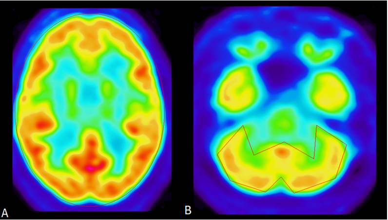
Figure 1 PET images of Neocortical to Cerebellum Region of Interest (ROI) analysis. Defendant has decreased neocortex to cerebellar ratio compared to normal controls consistent with someone who has sustained multiple traumatic brain injuries causing diffuse decrease in neocortex relative to the cerebellum. Seen in patients at high risk for CTE such as ex-NFL football players. A, The mean neocortex for ROIs is 1.00 whereas controls averaged 0.97, standard deviation=0.01, z-score= 2.21, p-score = 2.7E-02. B, The mean cerebellum for ROIs is 1.11 whereas controls averaged 0.94, standard deviation=0.07, z-score= 2.35, p-score = 1.9E-02.
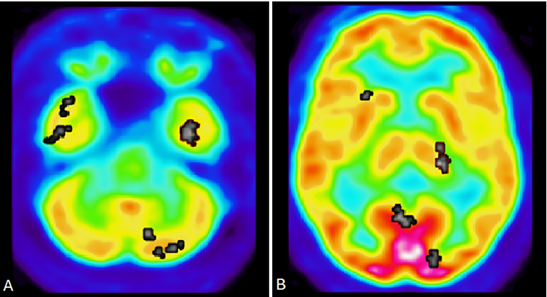
Figure 2 PET images of Frontal to Occipital Lobe Region of Interest (ROI) analysis. The mean frontal lobe/occipital lobe ratio for ROIs is 0.80 whereas controls averaged 0.94, standard deviation=0.06, z-score= -2.24, p-score = 2.5E-02. A, The mean frontal lobe for ROIs is 1.01 whereas controls averaged 1.02, standard deviation=0.04, z-score= -0.28, p-score = 7.8E-02. Defendant’s frontal lobe is especially compromised. The frontal lobe is involved with impulse control and judgment. B, The mean occipital lobe for ROIs is 1.25 whereas controls averaged 1.08, standard deviation=0.05, z-score= 3.17, p-score = 1.5E-03. He showed decrease frontal lobe metabolism relative to occipital lobe when compared to normal controls.
He also showed an abnormal increase in metabolism in the left inferior temporal gyrus with a metabolic value of 1.13 whereas controls averaged 0.89, standard deviation of 0.07, z score =3.72, p-score =204E-04 (Figure 3a). There was also an abnormal increase in left thalamus metabolism with metabolic value of 1.07 whereas controls averaged 0.82, standard deviation =0.05, z-score = 5.02, p-score =5.1E-07 (Figure 3b). These findings are consistent neurological consequences for severe emotional abuse that affect the developing limbic system from repeated stress.40 These PET findings of increased metabolism in left inferior temporal gyrus and left thalamus would also be consistent with the quantitative volumetric abnormalities reported below with decreased left forebrain parenchyma and asymmetrically decreased left sided hippocampus and amygdala since chronic seizures can result in brain atrophy.
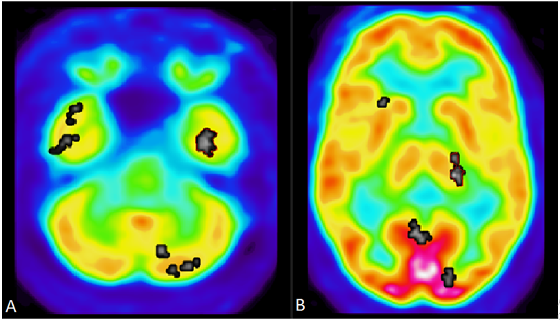
Figure 3 A, PET data shows abnormal increase in metabolism in the left inferior temporal gyrus (Brodmann area 20). B, PET data shows an abnormal increase in left thalamus metabolism. Findings are consistent with limbic kindling which has been proposed as a neurological consequence of severe emotional abuse when the developing limbic system is kindled by repeated exposure to stress.
Literature from Teicher and others have found evidence of spike and sharp wave EEG abnormalities in individuals with history of childhood abuse or neglect.41-43 These “limbic irritability” symptoms were similar to clinical and EEG findings seen in patients with temporal lobe epilepsy. Limbic kindling would result in abnormal metabolic activation of regions such as the left inferior temporal gyrus.44 The metabolic activation of the temporal gyrus would also result in downstream activation of the thalamus and would be consistent with the cortico-thalamo-pallidal-cortical loops.45 Limbic kindling due to emotional abuse results in abnormal activation of left inferior temporal gyrus consistent with epilepsy spectrum disorder, temporal lobe epilepsy, and with non-convulsive seizures with EEG and metabolic correlates of seizures without convulsions.46,47
There is an extensive body of medical literature from 1880 to 2013 relating epilepsy with violence and aggression which was reviewed by Pandya. Cases of epileptic patients who committed murder found 80% of the focal epilepsy had temporal epilepsy.48 In these cases the ferocious and excessive nature of violence is exhibited by multiple stabbings and more injuries than required to kill the victim. The nature of the stabbings in the second murder were consistent with this pattern of aggression reported in patients with epilepsy. Violence is associated with epilepsy even in the interictal phase and these episodes of violence were typically precipitated by situations of emotional salience which led to a period of built up agitation that then triggered an outburst of prolonged aggression lasting several minutes or longer.49,50 In this case, the emotionally salient trigger was the theft of the honey bun.
MRI-DTI analysis
Brain imaging assessments done with MRI Diffusion Tensor Imaging show significant abnormalities consistent with traumatic brain injury and high risk of chronic traumatic encephalopathy (Figure 4a). MRI-DTI findings found that the defendant displays very significant abnormal decreases in FA in the left anterior corpus callosum (mean FA ROI ratio is 0.37 whereas controls averaged 0.58, standard deviation=0.04, z-score= -5.38, p-score =, 7.6E-08). The decrease in FA in the mid corpus callosum is consistent with significant and multiple TBIs received in the past. He also has very significant abnormal decreases in the left internal capsule FA (mean FA is 0.25 whereas controls averaged 0.42, standard deviation = 0.02, z-score =-7.91, p-score =2.5E-15 (Figure 4b). The odds of this occurring by chance alone are 2.5 out of quadrillion.
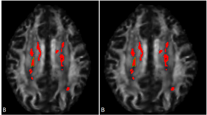
Figure 4A, MRI-DTI findings found that the defendant displays very significant abnormal decreases in FA in the left anterior corpus callosum. B, Diffusion Tensor Imaging (DTI) Statistical Analysis. Left anterior corpus callosum and left internal capsule display high risk of CTE. Abnormal decrease in fractional anisotropy (FA) in left internal capsule consistent with traumatic brain injury. P-value 2.5E-15, indicated the chances of this occurring by chance is 2.5 out of a quadrillion.
Tractographic analysis shows that there is an asymmetrical decrease in fiber track length in the left middle corpus callosum compared to the right (Figure 5a) Decreased track fibers were found in DTI Tractography in the left hemisphere of the corpus callosum compared to the right hemisphere (Figure 5b). These DTI findings are predominantly left sided and would be consistent with left sided head trauma. They would also be consistent with his history of having been beaten in the head repeatedly by a right handed individual such as his abusive mother growing up. They would also be consistent with left side limbic kindling noted above in the PET scan analysis.
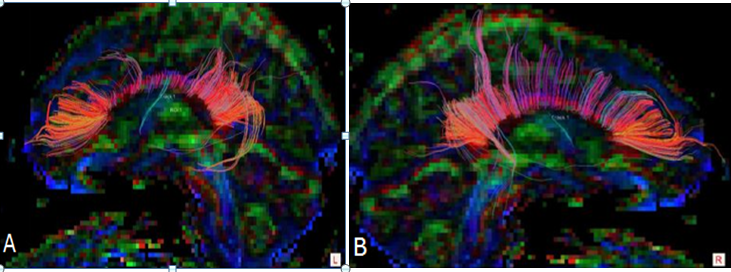
Figure 5A DTI-Tractography images in Corpus Colosseum, white fibers tract in left hemisphere is abnormal than right hemisphere. B Decreased fiber track length on the left side of the corpus callosum compared to the right side show lack of connectivity. Tractography suggest defendant for high risk of CTE and even Alzheimer’s disease.
Other MRI findings
There are punctate T2 and inversion recovery hyperintensities in the left frontal periventricular white matter identified. These are nonspecific but can be seen in cases of axonal shearing from brain trauma as well as microvascular disease. There are no focal or diffuse signal abnormalities on the axial DWI imaging (Figure 6). The SWI-venous bold images are remarkable for a punctate area of signal abnormality in the left frontal periventricular white matter which may reflect blood product and/or hemosiderin deposition. The DTI- Tractography images demonstrate some thinning of the corona radiata fiber tracts bilaterally.
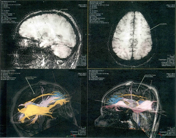
Figure 6 DTI- Tractography images demonstrate some thinning of the corona radiata fiber tracts bilaterally.
MRI brain quantitative volume analysis
Statistical analysis of NeuroQuant volumetric measures of MRI data shows a significant reduction in left and right forebrain parenchyma and left and right cortical gray matter in absolute and relative terms, which is more pronounced in relative terms on the right side. The defendant shows significant enlargement in the right lateral ventricle in relative terms. He shows a reduction in the left hippocampus, left amygdala, and left caudate and right putamen in absolute terms. He shows a bilateral reduction in the thalamus and cerebellum in absolute terms. Data shows a significant reduction in intracerebral volume in absolute terms. These findings are consistent with damage from premature birth, TBI, and severe childhood emotional abuse. The predominantly left sided nature of the atrophy noted in brain structures such as the hippocampus and the amygdala would be consistent atrophy secondary to multiple traumatic brain injuries from having been beaten in the head repeatedly by his abusive mother when he was growing up.
From Quantitative Volumetric analysis, the defendant shows significantly lower brain volume compared to normal, very significant reduction in left and right forebrain parenchyma, which is more pronounced on the left than the right. The reduction in his left forebrain parenchyma is more than six standard deviations below the mean. That means these findings would be seen in only 8.7 out of ten billion people. The quantitative volumetric analysis also shows left-sided asymmetrical decreases in the hippocampus and amygdala. Left-sided atrophy to the hippocampus and amygdala would be consistent with being hit on the head multiple times by a right-handed person. The reduction in left forebrain parenchyma and the left sided asymmetrical decrease in hippocampus and the amygdala would also be consistent with atrophy secondary to chronic temporal lobe epilepsy on the left side. This left-sided reduction would also be consistent with the abnormal increase in left inferior temporal lobe metabolism and left thalamus (see above PET discussion), and with abnormal decrease in FA in the left internal capsule, left anterior corpus callosum (see above DTI Discussion). This left-sided reduction would also be consistent with an asymmetrical decrease in fiber track length of the left mid corpus callosum compared to the right side.
Neuroquant general and triage brain atrophy report
NeuroQuant General and Triage Brain Atrophy Reports show multiple cortical sub-regions were in the 1st percentile including Intracranial Volume (ICV) of 1384.76 cm3 (Table 1). Multiple brain regions were noted to be in the 1st percentile on the Brain Triage atrophy report (Table 2).
Imaging supports neurological dysfunction
Providing neuroimaging from MRI-DTI, MRI quantitative volumetric, and PET scan evidence for the capital mitigation case supports the defendant's conditions of neurological consequences of childhood emotional and physical abuse such as limbic kindling, TBI, and high risk of developing CTE. From the psychological examination, the defendant displays history and symptoms consistent with pre-existing head trauma, emotional trauma, and psychological trauma from his early upbringing. The neuroimaging and the psychological assessment provides testimonial evidence of the defendant's significant abnormalities in developing neurological sequelae from physical and emotional abuse such as limbic kindling, and multiple TBIs with the high risk of CTE. Testimonial evidence also found the decreases in FA from the left anterior corpus callosum. The defendant’s QV displays lower brain volume than normal consistent to left-side atrophy.
Advantage of MRI-DTI and PET
Water diffusion is typically random and the same in all directions in an unconstrained environment. MRI-DTI and fiber Tractography tract white matter fiber to analyze human brain abnormalities by looking at water diffusion51-53 to determine whether water diffusion is hindered or constrained from diffusing in directions perpendicular to the white matter fiber or has higher diffusion in directions parallel to fiber in the intact axon. The directionality of the water diffusion is called fractional anisotropy and the fractional anisotropy is higher in an intact axon than a typical damaged axon. In a damaged axon which may be sheared, the water molecule can more easily diffuse through the walls of the damaged axon in a direction perpendicular to the axon. Diffusion tensor imaging is considered more sensitive and robust at detecting signs of traumatic brain injury than former MRI sequences.54 FA is given a value of 0 to 1; higher values indicate more white matter tract integrity.55 Typically, FA for healthy corpus callosum is .75 + .05 as FA for TBI is .60, which would be 3 SD below mean, p = .001.56
PET metabolic imaging is more sensitive for diagnosing TBI and psychosis than conventional MRI structural imaging. PET scans have been utilized frequently in court trials, providing a visual representation of regional brain function. As electrons are negative, positive electrons are a form of antimatter that are created by a cyclotron and attached to a tracer of sugar metabolism and then injected into patients. When positrons combine with electrons, a matter-antimatter conversion is formed to release tremendous energy; two gamma rays emerge that are 180 degree apart and detected by crystal detectors in the PET camera.57, 58 Thus, use of these techniques further reveal the defendant’s display of traumatic axon shearing injury, which diffused axon injury can cause shortening.
CTE
CTE is linked to accumulation of multiple TBIs due to neurodegenerative changes hyperphosphorylated tau and TDP-43 protein.59-61 As previously mentioned, results lead to a progressive decline of memory and cognition, poor impulse control, and aggressiveness. Early stages of TBI, before age 12, increases the risk of chronic traumatic encephalopathy as seen in ex-NFL football players and combat military veterans.62 His traumatic history may have shown early signs of TBI when he was beaten frequently in the head and tied to a ceiba tree by his abusive mother. Adolescents with multiple concussions may be more susceptible to diffuse brain injury, which leads to more pronounced and prolonged cognitive deficits.63
Childhood abuse effects
The defendant experienced significant childhood abuse. These abuses not only involved his mother’s abusive treatment but also included his grandmother as well; who starved him severely as a form of punishment requiring him to have to obtain food from outside the home. The defendant’s capacity to conform to the law requirements is largely impaired by neurological damage, emotional abuse, and psychological cruelty.
Exposure of physical and emotional abuse from parents during childhood is a factor that can contribute to posttraumatic stress disorder in adults. 64 Like CTE, PTSD can damage impulse control centers such as the amygdala and increase the risk of developing impulse disorders. Patients with brain injury develop depression and can have irrational aggressive behavior.65 Forms of domestic violence described as “a type of war zone” from childhood experiences can predict what they perceive as attacks.66 This explains the defendant’s aggression towards the inmate he stabbed when confronted in a closed cell. Overall, brain injuries increase the risk of other neuropsychiatric complications.
Mental retardation is not the cause for the finding
Although the patient was considered mentally retarded according to neuropsychological testing, his imaging abnormalities are not consistent with what has been reported for mental retardation. The FA score from the MRI DTI for the corpus callosum has been reported to be significantly lower than those of normal intelligence.67,68 While the decrease is significant (P<.01), the difference in FA is 3.8% between normal controls and the mentally retarded; whereas, the patient’s FA score is 40% lower than that of normal controls. This difference far exceeds the difference due to mental retardation and is much more consistent with multiple traumatic brain injuries.
Discussion
We report a case of Raul Roque, who is a 60-year-old convicted felon. This case report demonstrated the respondent’s traumatic childhood experiences that negatively led to and impacted the development of his cerebrum. The defendant’s severe emotional and physical abuse from his childhood significantly affected neurological changes. These neurological changes led to behavioral outbursts of prolonged aggression and devastatingly, violence; committing homicides on two occasions within ten years apart. PET, MRI-DTI, quantitative volumetrics, and psychological examination align with all the defendant’s family members' depositions and stories. Through brain image data analysis, the repeat offender shows to be at high risk for chronic traumatic encephalopathy, multiple traumatic brain injuries, and post-traumatic stress disorder from severe childhood negligence in Cuba. As the quality and precision of neuroimaging advances, it not only serves a purpose for research, yet can also be a beneficial tool for mitigation in the court of law, which expanse litigations to use science as another supplemental of evidence towards criminals who are facing death penalty. Neuroimaging fills in the gaps of what the litigations standpoint is for death penalty. Our findings not only helped the obtain prognosis but assist detainees who face death penalty to have a fair shot in life again, despite behind bars.
Neuroscientific advances in brain imaging enable objective and quantitative assessments of multiple dimensions such as regional cerebral metabolism with PET, regional assessment of the integrity of white matter tracts with DTI, and regional atrophy with quantitative volumetrics. These advances can be used to provide corroborative evidence of significant early childhood abuse and neglect which alters brain function and structure, and affects the ability to regulate aggressive behavior. Neuroscientific informatics are already utilized for primary research, clinical practice, and medical aspects, but are now being increasingly utilized in law; hence, neurolaw.69-71 Despite the recent advancements in neurolaw, many argue that neurological evidence of cognitive impairments, diseases, and disorders mitigates culpability of the crime committed; especially in murder cases.72 Although there is strong opposition to the interpretation and utilization of neurological evidence, Florida State’s trial is one example of how neuroimaging from expert testimonials can help mitigate a death penalty case. Brain imaging techniques and other neurolaw principles can be used in homicide court trials to detect neurobiological abnormalities resulting from physical and emotional trauma in murderers.
None.
None.

©2022 Go, et al. This is an open access article distributed under the terms of the, which permits unrestricted use, distribution, and build upon your work non-commercially.