eISSN: 2473-0815


Case Report Volume 12 Issue 2
MD, Professor of Potiguar University, Brazil
Correspondence: Tabata de Alcantara, MD, Professor of Potiguar University-Brazil.
Received: June 24, 2024 | Published: July 10, 2024
Citation: de Souza TVP, Ferreira TA, de Oliveira HFV, et al. Pamidronate and fassier-duval telescopic stem: pillars in the treatment of osteogenesis imperfecta. Endocrinol Metab Int J. 2024;12(2):52-53. DOI: 10.15406/emij.2024.12.00347
Osteogenesis imperfecta is a systemic genetic disease of connective tissue with a prevalence of 6 to 7 per 100,000 births, affecting collagen type 1 containing tissues, especially bone tissue. Low bone mass is its main characteristic, which causes fragile bones, susceptible to deformities and recurrent fractures.1 Approximately 90% of individuals are heterozygous for mutations in the COL1A1 and COL1A2 genes, with a dominant inheritance pattern or sporadic mutations.2
Fractures can occur at any stage of life, but mainly in childhood. In some children, the first ones starts when they begin to walk, because the upright posture increases the weight load on the lower limbs.1
The first descriptions of the use of pamidronate in the treatment of severe osteogenesis imperfecta occurred in 1998 and its major benefits are a reduction in the number of fractures, increase the muscle mass and in the growth speed.3 Along with bisphosphonates, the use of the Fassier-Duval telescopic intramedullary nail is a pillar in treatment, providing the possibility of correcting deformities and reducing fractures.
We evaluated the case of a patient diagnosed with Osteogenesis imperfecta type 3 according to the Sillence classification. E.M.B., a six-year-old male, has been treated with pamidronate since he was two years old, at a dose of 1 mg/kg/day for three days every four months. Before starting the medication, he had been admitted to the emergency department 14 times for fractures.
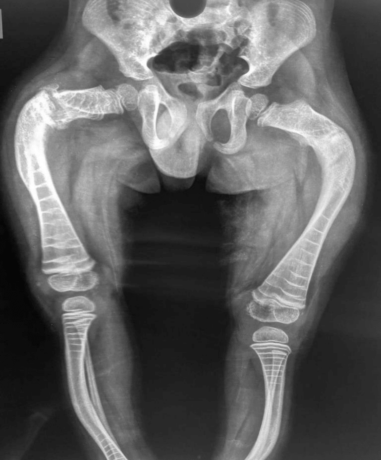
Due to the frequent lower limb fractures and hospital admissions, it was indicated surgical treatment of deformities through osteotomies and fixation with telescopic intramedullary nail. (Figure 1)

Figure 1 Pre-operative X rays where we can see the deformities in anterior and lateral plane of the femur and tíbia.
He was operated for correction of right and left femur on a same surgical time in March 2022. Posteriorly he was underwent right tibia treatment in august 2022 and left tibia in May 2023. (Figures 2)
His progress was extremely satisfactory, with no fractures occurring in the respective long bones subjected to the procedure after placement of the rods. Before their placement, the average number of fractures was six per year. Bone growth was maintained and the patient is now able to walk at home with devices.
Osteogenesis imperfecta is a genetic connective tissue disease that, in 70% of individuals, is caused by mutations in one of two COL1A1 and COL1A2 genes that code for type I collagen chains.3 The clinical presentation is extremely variable, including increased susceptibility to fractures, reduced bone mass, short stature, progressive skeletal deformities, bluish sclera, dentinogenesis imperfecta, ligament laxity and hearing loss.4
According to Sillence's classification based on clinical and radiographic evaluation, there are subtypes of osteogenesis imperfecta. Type 1 is the most common, with bluish sclera and a good prognosis; type 2 is a lethal perinatal form, but which currently survive with proper care, is a more severe form of the disease; type 3 is severe
with progressive deformities and normal sclera, here the triangular face and the multiple fractures at the birth are common; and type 4, also with normal sclera, is intermediate in severity, children have fractures until adolescence, decreasing in frequency. There is also type 5, a moderate form of the disorder, with distinct characteristics, such as an interosseous membrane between the radius and ulna.1 Acquisitions of genotypic analysis and variations of the original types were added during the years.5,6
Our patient corresponds to type 3, which benefited most from the use of pamidronate as well as telescopic intramedullary nails, as it corresponds to the most severe non-perinatal lethal type 2.
The medical and surgical treatment of osteogenesis imperfecta has undergone two revolutions that have improved quality of life and functional capacity: reduced bone absorption with the use of bisphosphonates and improved internal fixation with the development of the Fassier-Duval telescopic rod.7
Pamidronate infusion was the standard treatment, but zoledronic acid is increasingly used to treat children with osteogenesis imperfects.8
The placement of the rod is indicated in patients who are old enough to walk, have axial deformity greater than 20º and have more than two fractures in the same bone in one year. The telescopic rod provides 48% greater survival at four years, as well as a significant improvement in gait and autonomy.5 An important advantage of this technique is that several bones can be treated during the same surgical procedure and this reduces rehabilitation time.3,9
In our study, the patient had an average of six fractures per year before underwent placement of rods in the lower limbs and there were no reports of any fractures after the surgeries, corroborating the great advantage of this technique, a reduction in the possibility of fractures with a consequent increase in survival.
There is no description of an appropriate time to start treatment with bisphosphonates. It is indicated after diagnosis and has been successfully performed in children under three months of age. Its use aims to increase bone mineralization, with a consequent reduction in the number of fractures and improved quality of life.4 This treatment permits patients with better and bigger bones, facilitating surgical interventions.
Pamidronate increases bone mass, reduces musculoskeletal pain, increases the height of the vertebral body and reduces the frequency of fractures.4
This patient started its use at two years old, and had almost 6 fractures of long bones per year during treatment, showing that only this intervention is not sufficient. Complete the treatment with roods was essential to decline the fractures numbers and permit efficient rehabilitation.
The current mainstays of treatment for osteogenesis imperfecta are the use of bisphosphonates and the placement of Fassier-Duval rods. Although pamidronate increases bone density, it has no effect on the femoral and tibial deformity present in the most severe children.3
Telescopic intramedullary nails by combine correction of bone deformity combined and bone stabilization have proven to be the most effective treatment for preventing fractures in the long term, as well as reducing the possibility of bone deformities, improving locomotion and rehabilitation. It should always be remembered that the treatment of this condition should always be multidisciplinary.
None.
The authors declare there are no conflicts of interest.
None.

©2024 de, et al. This is an open access article distributed under the terms of the, which permits unrestricted use, distribution, and build upon your work non-commercially.