Advances in
eISSN: 2572-8490


Research Article Volume 2 Issue 3
Department of Conservative Dentistry and Endodontics, Vydehi Institute of Dental Sciences and Research Centre, India
Correspondence: Kanika Varshneya, Department of Conservative dentistry and Endodontics, Vydehi Institute of Dental Sciences and Research Centre, Bangalore, Karnataka, India, Tel 9901064313, Fax 080-28416199
Received: February 19, 2017 | Published: May 8, 2017
Citation: Varshneya K, Benjamin S, Naveen DN, et al. Microleakage in class V cavities restored with composite resin using chitosan and consepsis as the cavity disinfectants. Adv Tissue Eng Regen Med Open Access. 2017;2(3):176–180. DOI: 10.15406/atroa.2017.02.00029
Aim: To compare the influence of chitosan and consepsis on bonding of composite resin to dentin and cementum.
Materials and methods: Class V cavities were prepared on 20 extracted mandibular molars. The cavities in the experimental groups were pre-treated with a scrub of cavity disinfectants followed by application of bonding agent. Preparations without cavity disinfectants served as control. After the cavities were restored with resin composite (G-Aenial Universal Flo), the specimens were subjected to dye penetration. Statistical analysis was performed using Chi-square test and Fisher Exact test.
Results: There was no statistically significant difference in the leakage values between the Chitosan and Consepsis group. There was a statistically significant difference between the microleakage score of the bonding system with or without the cavity disinfectant.
Conclusion: Chitosan was found to be similar to Consepsis, an established cavity disinfectant in preventing microleakage and was found not to interfere with bonding of composite resin to either dentin or cementum, holds potential to be used as an effective cavity disinfectant prior to restoration with composite resin.
Keywords: chitosan, cavity disinfectant, chlorhexidine gluconate, dentin-bonding resin, microleakage
CEJ, cemento enamel junction; NaOCl, sodium hypochlorite; CHX, chlorhexidine
The setting reaction of resin composites involves polymerization shrinkage that may lead to the formation of a contraction gap at the tooth restoration interface. This gap can result in the passage of bacteria, fluids, or ions between the cavity wall and the resin composite, a process which is known as microleakage.1 Previously, many new bonding systems have been introduced to reduce the size and incidence of gap formation following placement of a resin composite restoration. Even then, microleakage, especially at the dentin (cementum) aspect of restoration, remains a problem of clinical significance.2–4 Microleakage has been demonstrated as a factor in hypersensitivity, secondary caries and pulpal pathology.5
The success of the restorative procedures depends on the effective removal of infected dentin, prior to the placement of the restorative material. The main problem associated with microleakage can be magnified by residual caries, as a consequence of failure to mechanically remove the infected tooth structure.6 Histological and bacteriologic studies have shown that very few teeth are actually sterile after cavity preparation and that bacteria left in the cavity preparation could survive for longer than a year after removal of the carious dentin, it is therefore important to eliminate any remaining bacteria that may be left behind in the smear layer, at the enamel-dentin junction, or in the dentinal tubules.7 Thus, application of disinfectants after cavity preparation and before restoration is fast gaining acceptance. It eliminates risks due to bacterial activity.8,9 However, there is concern about the interference of cavity disinfectants with dentin bonding agents, since they have been shown to alter the sealing ability of the hydrophilic resin to the dentin.10 Contrary to this concern, has been a suggestion that cavity disinfectants can improve the sealing ability of dentin bonding agents by rehydrating the conditioned dentin. The purpose of this study was to evaluate the effect of two cavity disinfectants on the microleakage of a non-rinse dentin-bonding system, Clearfil SE Bond (Kuraray).
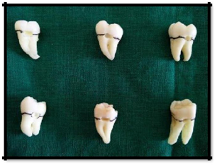
Twenty freshly extracted human mandibular molars, free of cracks, caries and restorations, were used in the study. The teeth were scraped of any tissue remnants and stored in 2.6% NaOCl (Sodium hypochlorite) solution (VIP, Vensons India, Bangalore, India) for 15minutes each and rinsed under running water. They were later cleaned with pumice and stored in normal saline (NS, Fresenius Kabi, Goa, India) at 40˚C until use (Figure 1).
Standardized class V cavity preparations were made on the facial and lingual surfaces of each tooth, with no 245 straight fissure bur (Mani, India) in a high speed hand piece (NSK Pana Air, Japan) utilizing water-spray coolant. Standardized preparations were obtained by making cavity preparations approximately 2mm wide, 2mm deep and 3mm long, paralleling the cemento-enamel junction (CEJ). The depth of the preparations was assessed using a periodontal probe (GDC, India). The gingival halves of the preparation were extended 1mm below the CEJ (Figure 2). Prepared surfaces were rinsed with distilled water for 20seconds followed by air drying for 20seconds a two-way syringe. The teeth were then randomly divided into two groups:

Figure 2 Standardized class V cavity preparations were made on the facial and lingual surfaces of each tooth.
Group I consisted of ten teeth (20 cavity preparations);10 cavity preparations were treated with CHX (chlorhexidine) based cavity disinfectant solution (Consepsis, Ultradent USA), followed by the application of a dentine bonding system (Clearfil SE Bond, Kuraray, Japan). Remaining 10 cavities were bonded without chlorhexidine pre-treatment and were used as control.
Group II consisted of ten teeth (20 cavity preparations);10 cavity preparations were treated with chitosan based cavity disinfectant solution (Everest Biotech, Bangalore), followed by the application of a dentine bonding system (Clearfil SE Bond, Kuraray, Japan). Remaining 10 cavities were the control i.e. without application of chitosan.
In each of the experimental groups, cavity disinfectant was applied with a sterile brush applicator (Dochem, Shanghai, China) and scrubbed for 20seconds; excess disinfectant was removed by lightly air drying for five seconds.
After cavity disinfection, the dentin bonding system (Clearfil SE Bond, Kuraray, Japan) was applied as per the manufacturer’s instructions. Primer was applied to the cavity floor and agitated for 20seconds and gently air dried. A layer of bonding resin was applied to the preparation with a brush, spread gently with air and cured for 10seconds. The cavity preparations were restored with a resin composite (G-Aenial Universal Flo, GC, Tokyo, Japan) by light curing for 60seconds. The cavosurface margins were then finished with a finishing bur and 3M USA discs. All the teeth were stored in distilled water for 24hours, at 37˚C, and subjected to 1,000 thermal cycles between water baths of 50˚C and 550˚C, with a dwell time of 30seconds. The teeth were then subjected to dye leakage tests.
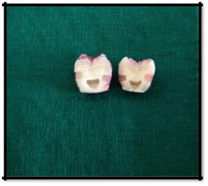
All the specimens were covered with two coats of nail varnish leaving 1 mm of the tooth-restoration margin and the root apices were sealed with modelling wax. The specimens were then immersed in 0.5% basic fuchsin dye (NICE Chemicals Pvt Ltd, India), in separate sealable glass vials (Borosil, India), at 37˚C for 24hours. After staining, the teeth were rinsed in water and dried using two-way syringe, the radicular parts of the teeth were then cut 4.5mm below the CEJ using diamond discs. Coronal portion of the teeth were sectioned buccolingually, in the approximate centre of the restoration (Figure 3).

Figure 3 Coronal portion of the teeth were sectioned buccolingually, in the approximate centre of the restoration.
Microleakage was assessed for both occlusal (enamel) and gingival (cementum) margins, using a stereomicroscope (Magnus) at original magnification of X16.
The depth of the stain (dye leakage) was judged according to the following scale:
Results
Control group showed consistently higher leakage values ranging from 3 to 4 whereas the experimental groups exhibited significantly less leakage (0, 1, 2) (Table 1) (Table 2). Chi-square/Fisher Exact test was used for the significance of study parameters on categorical scale between the groups.
Leakage Pattern in Experimental Groups |
Group A |
Group B |
No leakage |
3(30.0%) |
6(60.0%) |
Penetration less than on half of the length of occlusal / gingival wall |
3(30.0%) |
4(40.0%) |
Penetration greater than on half of the length of occlusal / gingival wall |
2(20.0%) |
0 |
Penetration up to and along the axial wall |
2(20.0%) |
0 |
Penetration within the pulp |
0 |
0 |
Total |
10(100.0%) |
10(100.0%) |
Table 1 Comparison of Leakage pattern in two groups in experiment.
P: 0.204; Not significant; Fisher Exact test
Leakage Pattern in Control Groups |
Group A |
Group B |
No leakage |
0 |
0 |
Penetration less than on half of the length of occlusal / gingival wall |
0 |
0 |
Penetration greater than on half of the length of occlusal / gingival wall |
1(10.0%) |
1(10.0%) |
Penetration up to and along the axial wall |
4(40.0%) |
5(50.0%) |
Penetration within the pulp |
5(50.0%) |
4(40.0%) |
Total |
10(100.0%) |
10(100.0%) |
Table 2 Comparison of Leakage pattern in two groups in Controls.
P: 0.206; Not significant; Fisher Exact test
The scores for leakage along the occlusal and gingival margins in the treatment groups were compared using Wilcoxon matched pairs signed rank test. The level of significance was established as P<0.05, for all the tests. Pair wise comparisons for leakage amongst the two experimental groups in both the occlusal and gingival walls showed no statistically significant differences as shown in Graph 1. However, a statistically significant difference was found between both the experimental groups and their respective controls (Graph 2).
Discussion
Historically, it was suggested that dentin should be sterilized before the placement of any restorative material. Many chemicals, such as silver nitrate precipitated with eugenol, thymol, and potassium ferrocyanide, had been proposed for this purpose. The rationale prevailing for this was that any residual microorganisms should be eliminated in order to prevent the potential propagation of caries. Today, it is known that these chemicals are irritating to the pulp when applied to the dentin surface.11 Thus, any chemical that is capable of destroying microorganisms may also have a detrimental influence on the pulp. The use of consepsis and chitosan-based disinfectant has been proposed for disinfecting the cavity preparation, prior to its restoration.
A study by Perchyonok12 had reported that the antioxidant-chitosan hydrogels significantly improved bonding to dentine with or without phosphoric acid treatment. The Chitosan used in our study is a natural carbohydrate polymer derived from the deacetylation of chitin produced commercially from crab and shrimp shell wastes. Numerous studies have shown that chitosan is a biologically safe biopolymer, has been proposed as a bio-adhesive polymer13 Most of the current generation disinfectants contain 2% chlorhexidine gluconate as the primary active ingredient, which is an antiseptic with a wide spectrum of action. Consepsis used in our study contains 2% chlorhexidine gluconate and has been reported to have better antimicrobial activity.
According to Shafiei et al.14 CHX acts as a preservative on dentin bonding and showed no adverse effect on immediate bond strength and enamel or dentin leakage. The use of cavity disinfectants after tooth preparation and before the application of dentin-bonding agents could help reduce the potential for residual caries and postoperative sensitivity.15 However, any positive benefits would be negated if the solutions significantly increased the amount of microleakage, by interfering with the bonding agent’s interaction with dentin.
In comparison to the control, both the experimental groups exhibited significantly lower levels of microleakage (Table 3) (Table 4). In the present study, Chitosan showed marginally better resistance to microleakage as compared to consepsis. This could be because of the formation of complexes between chitosan and metal ions in the inorganic dentin which is most probably due to the mechanisms of adsorption, ion exchange and chelation.12 Also, 0.12% and 0.25% (w/w) chitosan does not adversely affect adhesive properties of the bonding system.16
Leakage Pattern in Group A |
Experimental |
Controls |
No leakage |
3(30.0%) |
0 |
Penetration less than on half of the length of occlusal / gingival wall |
3(30.0%) |
0 |
Penetration greater than on half of the length of occlusal / gingival wall |
2(20.0%) |
1(10.0%) |
Penetration up to and along the axial wall |
2(20.0%) |
4(40.0%) |
Penetration within the pulp |
0 |
5(50.0%) |
Total |
10(100.0%) |
10(100.0%) |
Table 3 Comparison of Leakage pattern in two groups in Group A.
P: 0.013*; Significant; Fisher Exact test
Leakage Pattern in Group B |
Experimental |
Controls |
No leakage |
6(60.0%) |
0 |
Penetration less than on half of the length of occlusal / gingival wall |
4(40.0%) |
0 |
Penetration greater than on half of the length of occlusal / gingival wall |
0 |
1(10.0%) |
Penetration up to and along the axial wall |
0 |
5(50.0%) |
Penetration within the pulp |
0 |
4(40.0%) |
Total |
10(100.0%) |
10(100.0%) |
Table 4 Comparison of Leakage pattern in two groups in Group B.
P: 0.009**; Significant; Fisher Exact test
In our study, Consepsis solution also did not adversely affect the sealing ability of Clearfil SE bond.9,17 The presence of statistical difference in our study, between the control and the experimental groups treated with chitosan and consepsis before the application of the dentin adhesive systems, in both occlusal and gingival marginal leakage scores, showed that cavities treated without a cavity disinfectant had significant microleakage whereas Chitosan was found to be similar to Consepsis, an established cavity disinfectant in preventing microleakage and was found not to interfere with bonding of composite resin to either dentin or cementum, holds potential to be used as an effective cavity disinfectant prior to restoration with composite resin. The results of our study were in accordance with another study conducted by Meiers et al.9
Within the limitations of this study, it can be concluded that Chitosan was found to be similar to Consepsis, (an established cavity disinfectant) in preventing microleakage and was found not to affect the sealing ability of Clearfil SE Bond or interfere with bonding of composite resin to either dentin or cementum. Also, it holds potential to be used as an effective cavity disinfectant prior to restoration with composite resin.
The authors are thankful to Dr. Usha G, Mr. Bharat, Prime Dental, Ultradent, USA and Kurraray, Japan for providing cavity disinfectants and other materials related to the study.
The author declares no conflict of interest.

©2017 Varshneya, et al. This is an open access article distributed under the terms of the, which permits unrestricted use, distribution, and build upon your work non-commercially.