Advances in
eISSN: 2377-4290


Mini Review Volume 10 Issue 3
Department of Ophthalmology, Universitas Airlangga/Dr. Soetomo Hospital, Indonesia
Correspondence: Arantrinita Evelyn Komaratih, Department of Ophthalmology, Universitas Airlangga/Dr. Soetomo Hospital, Surabaya, Indonesia
Received: May 10, 2020 | Published: June 30, 2020
Citation: Komaratih AE. Early diagnosis glaucoma using spectral domain OCT. Adv Ophthalmol Vis Syst. 2020;10(3):65-68. DOI: 10.15406/aovs.2020.10.00387
Optical Coherence Tomography (OCT) is new technique that produces images of biological tissue by measuring the reflection of light from the structure being examined. OCT can greatly assist clinicians in diagnosing and managing glaucoma, especially early disease, when used in conjunction with serial clinical exams and visual field testing. Identification of structural damage to the optic nerve and retinal nerve fiber layer (RNFL) is an important component in the diagnosis of glaucoma. Spectral Domain OCT (SD-OCT), allows an earlier examination of these structures and is equipped with an analysis of the macula area to detect ganglion cell damage.
Keywords: early diagnosis, glaucoma, ONH, RNFL, GCC, GPA, OCT, spectral domain OCT, SD-OCT
Glaucoma is the third largest cause of blindness worldwide after cataracts and trachoma, reaching 14% of the total blind population. This causes glaucoma is the highest cause of irreversible blindness.1 Early diagnosis and glaucoma progression are needed so as to provide early treatment and slow progression. Glaucoma is an optic neuropathy disease caused by multi factors with the initial sign is structural retinal ganglion cell death then functional loss occurs in the form of a narrowing field of glaucoma characteristic, ending with irreversible blindness. These blindness complaints occur slowly; therefore glaucoma is called a vision thief. Optical Coherence Tomography (OCT) is a valuable diagnostic tool. The development of OCT technology has driven the success of glaucoma screening. Spectral domain (SD)-OCT technology has advantages over the previous generation of time domain (TD)-OCT in terms of increased scan speed and axial resolution so as to reduce distortion caused by eye movements2,3 With this ability, SD-OCT can analyze eye structures in more detail, especially in structures affected by glaucoma.
SD-OCT examination procedure
To obtain representative results, the OCT check must be carried out in the following way. First, enter the patient's identity and date of the examination correctly, this aims to make the data entered in accordance with the OCT normative data base and the results of the examination can be used to monitor progression periodically. Second, The pupils are dilated, the examination is done in dim light, and make sure the patient sits comfortably then the height of the OCT machine is adjusted so that the patient does not need to bend or sit too upright, aiming to prevent artifacts and make the examination process more comfortable. During the examination, the surface of the eye must not be dry (the patient is asked to blink for several times or given drops of artificial tears). Third, patients are asked to focus on the fixation point so that the scan can be centralized properly. If there is opacity on the visual axis, the patient may be asked to look a little further from the point of fixation or see an external fixation lamp. If the patient has poor fixation, then a better eye can be instructed to look at the external fixation lamp. The image acquisition circle is centered on the desired area and makes sure the image on the computer (viewing window) looks full structure. After correct placement, the image refocuses on the optic nerve area and fovea correctly, and then scanning can be done.
Assessing an OCT examination results obtained from signal strength, motion artifacts, and accurate acquisition. Poor signal strength can blur the boundaries between structures, so the quantitative results obtained are not correct. Excessive motion artifacts can cause data distortion, although in some OCT have been equipped with image tracking mode. Accurate acquisition is expected to be an image that is not cut and the placement of the image is right on the optic nerve and fovea.4
SD-OCT posterior segment examination in glaucoma
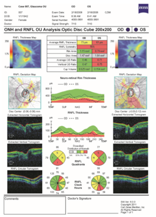
SD OCT examination in glaucoma consists of several components, including the analysis of the optic nerve head (ONH), retina nerve fiber layer (RNFL), and ganglion cell complex (GCC) in the macula. The ONH analysis of OCT is to assess the disc area, rim area, and cup-to-disc ratio. RNFL analysis was performed on average RNFL thickness, hemifield thickness, and quadrant RNFL thickness. Figure 1 shows printouts with measurements obtained from ONH and RNFL areas with one of the commercially available SD-OCT instruments, the Cirrus-OCT (Carl Zeiss Meditec, Dublin, CA, USA) OCT results of a 66-year-old woman (examination carried out in 2008) showed widening of the disc area, depletion of the rim area, and an increase in the vertical cup-to disc (C/D) ratio. RNFL analysis on average RNFL thickness obtained borderline thinning in the right eye and significant thinning in the left eye. Hemifield and quadrant RNFL thickness appear to be thinning in the Inferior, Superior, and Nasal regions of the right and left eye according to the ISNT rule typical of glaucoma.

Figure 1 ONH and RNFL OU analysis: optic disc cube 200x200.5
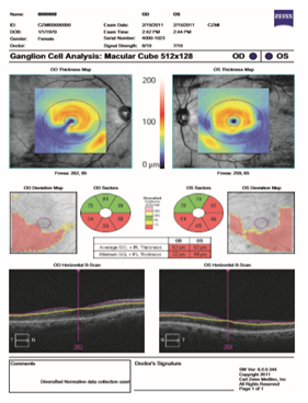
Then, in the macula region, an analysis of the ganglion cell complex is performed on average GCC thickness, hemifield GCC thickness, focal loss volume, and global loss volume. The measurement parameters are automatically compared with the normative database of OCT, and the results are color coded for "within normal limits" (green), "borderline" (yellow), and "outside normal limits" (red). Figure 2 shows a printout of ganglion cell analysis provided by a commercially available SD-OCT instrument the Cirrus-OCT (Carl Zeiss Meditec, Dublin, CA, USA) for a glaucomatous patient with decrease in average GCL + IPL thickness in all three inferior quadrants in the right eye and inferior and superotemporal quadrants in the left eye.

Figure 2 Ganglion cell analysis: macular cube 512x128.6
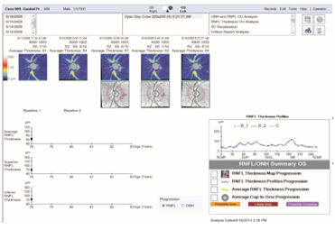
The results were collected to analyze the progression of the disease by assessing the linear regression trend on the ONH parameters and RNFL thickness starting from the beginning of the visit, with baselines obtained from measurements at the first 2 visits, then compared to the normative database variability. Interpretation of results is divided into three types namely "Possible Loss", "Likely Loss", or "Possible Increase" as shown in Fig.(3).

Figure 3 Guided Progression Analysis (GPA).7
Optical coherence tomography is effective in screening and early diagnosis of glaucoma in an office setting. This technology must be combined with subjective data, clinical examination results and other supporting examinations including tonometry, fundus photography, and visual field testing.
None.
None.
The authors have no affiliations with or involvement in any organization or entity with any financial interest (such as honoraria; educational grants; participation in speakers’ bureaus; membership, employment, consultancies, stock ownership, or other equity interest; and expert testimony or patent-licensing arrangements), or non-financial interest (such as personal or professional relationships, affiliations, knowledge or beliefs) in the subject matter or materials discussed in this manuscript.

©2020 Komaratih. This is an open access article distributed under the terms of the, which permits unrestricted use, distribution, and build upon your work non-commercially.