Advances in
eISSN: 2377-4290


Case Report Volume 4 Issue 3
Carlos Chagas Filho Biophysics Institute, Federal University of Rio de Janeiro, Brazil
Correspondence: Luiz Reis Barbosa Junior, Federal University of Rio de Janeiro-UFRJ, Rio de Janeiro, State of Rio de Janeiro, Brazil, Tel 55 21 96900-0027
Received: March 16, 2016 | Published: April 26, 2016
Citation: Reis L, Zangalli AL, Louzada RL, et al. Normative study of the full field electroretinogram in cats: a fast dark-adaptation curve. Adv Ophthalmol Vis Syst. 2016;4(3):66-69. DOI: 10.15406/aovs.2016.04.00107
Purpose: The aim of the present study is to standardize the normal values of each component of the signals that comprise the ERG of normal cats, in accordance with the ISCEV protocol currently recommended in clinical examinations, for its relevance as a diagnosis method to assess retinal function and detect lesions in their outer layers.
Methods: Ten healthy domestic cats were used for this study. The ERG was randomly recorded using a Nihon Kohden system. We registered a series of light and dark-adapted ERGs.
Results: The mean amplitude of the b wave in photopic conditions with bright white flash is 180,0 μV, while in scotopic environment we found a mean amplitude of 600,0 μV for the b wave, most of which was divided into two components (b1 and b2).
Conclusion: The cat ERG has a particular profile, with a fast dark-adaptation response. This normative study will allow the comparison of data recorded by different research groups, and enable a more complete use of the ERG as a tool for future studies on diagnosis and treatments of diseases affecting each retina layer in particular.
Keywords: electroretinogram, cat, standardization, dark-adaptation curve, retina
ERG: electroretinogram; ISCEV: enternational society for clinical electrophysiology of vision
The discovery of the electroretinogram (ERG) by Hoimgren (1865) was an important event in visual physiology that initiated the use of electrophysiology methods for studying visual systems. The Flash ERG offers the unique advantages of a readily recorded response that reveals electrical activity from certain class of retinal cells. It’s a mass response which is composed of elementary components arising from the different types of cells in the retina.1 The electroretinogram is a complex sequence of potential variation mainly studied by Granit since 1933. It is an electrical response produced by the retina, compared to a luminous stimulation. Granit recognized the necessity for separate analyses of ERGs from predominantly rod and pure cone retinas.
The three major components were designated PI, PII and PIII. In this terminology the term “P” stands for process, since at that time the cellular origin was not known for any of the potentials. The components were then numbered in the order in which they disappeared following the administration of ether anesthesia. After this procedure Granit found that the deflection of the cat ERG disappeared in three major steps. The c-wave disappeared first, and then the b-wave and off-response disappeared at about the same time, while the a-wave disappeared last.2 In 1954, Cobb et al.,3 demonstrated in the human ERG some small rhythmic waves, overlapping the upward phase of “b-wave”.
Yonemura, Masuda and Matta (1963) called the wavelets “the oscillatory potential”.4 The wavelets increase in amplitude with a stronger intensity of stimulating light, but are spaced at nearly equal interval of approximately 7 milliseconds with little regard to the intensity of stimulating light. Those observations have since been made on several animal species. The wavelets are designated in order of ocurrence as OP1, OP2, OP3 and so on. In 1989, the International Society for Clinical Electrophysiology of Vision (ISCEV) published the first internationally agreed standard for electroretinography. Standards for other common electrophysiological tests and for calibration of records equipment have been published more recently.5
Through this exam we evaluate the pathologies that affect retinal structures, sometimes without injured fundii. The ERG analyzes the electrical response of each retinal layer, can distinguish the response of each type of photoreceptor and can detect various defects on retinal cells, as well as, on experimental models of ocular diseases. It also assists in the diagnosis, management and monitoring of various diseases including: vascular occlusion, diabetic retinopathy, cone-rod dystrophies, retinal degenerations, and many others. We demonstrated in our study a standardization of electroretinography in cats and the response according to the time of adaptation in the dark light. All the waves of the electroretinogram will be studied.
Ten healthy domestic cats (felis catus) were used for this study, from both genders, aged between one and two years weighing two to three kg each. The animals were treated in adherence to the ARVO guideline for the care and use of animals in visual research. The ERG was randomly recorded from one eye of the previously anesthetized cat (intraperitoneal dose of ketamine hydrochloride and xylazine hydrochloride), using a Nihon Kohden system. The needle electrode was positioned at temporal limbus, the reference electrode was secured on the forehead and the ground electrode was attached at the ipsilateral leg. The electrodes were connected to a differential a.c. amplifier and the amplified signal was displayed by a direct oscilloscope. For procedures requiring a light stimulus of short duration a xenon strobo was used in intensity of 20 joules. The duration of the flash as specified by the manufacture was 10 usec. The full-field ERG records the electrical response of the retina potential after a light stimulus. In our laboratory we perform the full-field ERG with Nihon Kohden device and follow the standard protocol recommended by the International Society for Clinical Electrophysiology of Vision 2008 update.
The exam consists of five stages. In the first stage we evaluate the scotopic response of the rods after dark adaptation for 20 minutes and maximum pupil dilation, in order to assess the function of the rods by measuring the b-wave amplitude. The second stage of the examination is to assess the combination of the rod and cone system response, which evaluated the b-wave amplitude, generated by the Muller cells and indirectly by the activity of the bipolar cells; and the ratio of the wave b/a, studying the integrity of the inner nuclear layer. For the third stage, the dark adapted animal will have three measurements taken, all of which involve oscillatory potential, this shows the response of amacrine, interplexiform, and ganglion cells. During this stage of the experiment, the sum of the recorded amplitudes is taken, which allows us to evaluate the integrity of the microvasculature that supplies the inner retina. The fourth phase is the first photopic phase, and it occurs after adaptation to light for 10 minutes. This portion allows us to register the response of cones, while the suppression of the rods occurs. The last phase of the test consists in presenting high intensity flashes at a frequency of 30 Hz with the animal adapted to light. This allows us to evaluate the amplitude and the implied time of the wave, and thereby analyze the isolated integrity of the cone system.
Therefore, we registered for each animal a series of light and dark-adapted ERGs. The cat is light-adapted to suppress rod activity and a response is recorded to single flash of bright light. After that, the cats were adapted for 5, 10 and 20 minutes so as to record the scotopic ERG with both white and blue lights, as well as the isolation of the oscillatory potentials. The best results were found with five minutes of dark adaptation, generating waves with an acceptable standard and thereby are possible to optimize the tests using a less exposure time.
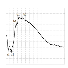
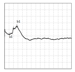
The mean amplitude of the b wave in photopic conditions (Figure 1) with bright white flash is 180,0 μV, while in scotopic environment (Figure 2), also with white flash, we found a mean amplitude of 600,0 μV for the b wave, most of which was divided into two components (b1 and b2). The largest signal found with a dark adapted retina is consistent with the fact of the cat’s retina being a rod predominant retina. Using a blue flash, the signal recorded mostly do not express the dichotomized a and b wave found with white flash. As the blue filter isolate the rod electrical activity, this result points out a cone origin of the first a1 and b1 components of the negative (a) and positive (b) deflections of the full field ERG (Figure 3). The oscillatory potential interpretation as the same of humans (Figure 4). We also analysis the c-wave in the ERG of cats, which shows the pigment epithelium function (Figure 5).

Figure 1 Scotopic ERG in cats. In a scotopic environment with white flash we found a mean amplitude of 600,0 μV for the b wave, most of which was divided into two components (b1 and b2).

Figure 2 Photopic ERG in cats. The mean amplitude of the b wave in photopic conditions with bright white flash is 180,0 μV.

Figure 3 Scotopic Blue Filter ERG of cats. We we used a blue flash, the signal recorded mostly do not express the dichotomized a and b wave found with white flash. As the blue filter isolate the rod electrical activity, this result points out a cone origin of the first a1 and b1 components of the negative (a) and positive (b) deflections of the full field ERG.
As long ago as 1865 it was discovered by Holmgren that the difference of electrical potential, already known since the work of Du Bois-Reymond (1848) to exist between the cornea and the back of the eye, altered when light fell upon the retina. This electrical response to illumination is now generally known as the electroretinogram. The time-course of the electroretinogram became accurately known almost as soon as sufficiently rapidly responding sensitive instruments were available to record it. Gotch (1903) determined its main features with the capillary electrometer, and von Brücker & Garten (1907) and Einthoven & Jolly (1908) established its details by means of the string galvanometer.
Comparatives studies shown that the main features of the electroretinogram are strikingly similar throughout all the classes of vertebrates. Illumination of the eye causes a cornea-negative a-wave, and then a cornea-positive b-wave. These are differently affected by the brightness of the light; for strong stimuli the may be about equal in amplitude, but weak stimuli can be found that produce large b-waves but no detectable a-waves. In the dark-adapted but not in the light-adapted eye the a- and b-waves are followed by a much slower c-wave, which is usually cornea-positive, but in the cat and rat (Parry, Tansley & Thomson, 1953) can be cornea-negative.
When the light is extinguished, in cold-blooded vertebrates, birds and a minority of mammals a cornea-positive response similar in its time-course to the b-wave is produced. This off-effect or d-wave is generally more prominent in the light-adapted than in the dark-adapted eye. The majority of mammals, including man, as well as the cats, differ from other vertebrates in that the d-wave is very small, and is sometimes cornea-negative. The best and influential analysis of electroretinogram is the latest one, that of Granit (1933), which recognizes three componentes, called PI, PII and PIII. The analysis was based on the effects of ether anaesthesia and of asphyxia. The changes in the electrical record during deepening anaesthesia were interpreted as due to the removal first of PI and then of PII, and those produced by asphyxia as due to selective removal of PII.
In mixed retinas, such as those of man and the cat, electroretinograms can be obtained in dark-adapted and light-adapted states, the spectral sensitivity changing with adaptation from that characteristic of rods to that characteristic of cones. Such diferences in form between the dark-adapted and light-adapted electroretinograms do not necessarily represent a change from rods to cones; for example, the electroretinogram of the cat has a fast dark-adaptation curve, its suggest that the degrees of dark-adaptation that are insufficient to convert the spectral sensitivity curve from the photopic to the scotopic stimuli.
The proper technique for looking for diferences between the electroretinograms provoked by activity of rods and of cones is to compare the responses of a mixed retina in a constant adaptational state to lights of diferente wavelength or, even better, diferente directions of incidence on the retina. For the light-adapted eye the responses produced by diferente colours are all brief and diphasic, a very small cornea-negative deflexion (a-wave) being followed by a larger cornea-positive one (b-wave). The response to deep red light is the same in the dark-adapted eye as inthe light-adapted, but with blue and green stimuli the brief diphasic response given by the light-adapted eye is replaced or masked, in the dark-adapted state, by a slower and much larger cornea-positive deflexion. The vertebrate retina contains three layers of nerve cells, the receptors, the bipolars, and the ganglion cells, joined to one another in a variety of combinations by cross connexions through the horizontal and amacrine cells. Not only is the anatomical structure of the retina very complicated, but is the anatomical structure of the retina very complicated, but so, as might be expected, are its physiological reactions. Stimulation of the eye by light, for instance, evokes several diferente types of response in the optica nerve as well as a complex retinal potential made up of at least three easily recognizable componentes not including the electrotonic potential along the optic nerve.5
The sensory elements (rods and cones) are connected to the bipolar cells which are in turn connected to the ganglion cells from which arise the optic nerve fibres. Convergence occurs by the connexion of several rods to one bipolar cell and of several bipolar cells to one ganglion cell. Cones are here represented as making individual connexions to their bipolar cells, but this condition is probably exceptional, occurring only in the human fóvea, not in the cats. The standard experiment on dark adaptation was introduced by Aubert in 1865 and consists-for work on visual sensation as well as on the electrical reactions of the eye-in measuring the fall of the threshold during a period in the dark. It will be seen that there are two phases of the fall in threshold. The eye was light adapted before dark adaptation was begun and it is, of course, essential that both the duration and intensity of the preliminar light adaptation should be strictly controlled.6
The dark adaptation of cats show a Purkinje shift in sensitivity. The red response is reminiscente of the dark adaptation curve of the human fóvea, the white response shows dark adaptation proper after an initial delay of nearly twenty minutes. The two lower curves illustrate characteristic variations in the course of dark adaptation of diferente isolated elements of the cat retina. In neither of these is there any initial delay, dark adaptation proper begins immediately. The eye adapts to either the light or in the dark situations. This adaptation is almost immediate if the luminance levels are close, it takes longer to settle, especially as regards the night vision, if the different light levels considerably from each other.7
The mechanisms that allow the dark adaptation are not strictly opposite of those that allow adaptation to light and to each other and possess their own limits. It is therefore necessary to consider separately and in succession: adaptation to darkness and light. We conclude that there must be two diferente types of rod, one reacting like cones so far as adaptation is concerned, the other like an “ideal” rod reflecting, fairly accurately, the course of visual purple regeneration. For instance, in the cat’s eye the b-wave may be so small as to be negligible after some minutes adaptation to 3.900 m.c., but no one with any experience of making visual purple extracts could possibly believe that the amount of visual purple present i salso negligible. On the other hand, the b-wave and visual purple regeneration both react in the same manner to changes in temperature.
The ERG is a mass response which is composed of elementar componentes arising from the diferente types of cells in the retina. Normal values were determined for the Flash ERG to both photopic and scotopic responses. However, it requires technical standardization for safety results. The cat ERG has a particular profile, with a fast dark-adaptation response. This normative study will allow the comparison of data recorded by different research groups, and enable a more complete use of the ERG as a tool for future studies on diagnosis and treatments of diseases affecting each retina layer in particular. We determined the time that it takes for the best response, and its obtained at 5 minutes of dark-adaptation, with a better final result. This strategy has the advantage of limiting the study of rod dark adaptation to a more reasonable amount of time, and it leads to a number that reflexts the kinetics of rod dark adaptation.
The author demonstrates that the standardization of the retinal electric reply in cats is necessary for future studies, as well as shows the particular retinal response with a fast dark-adaptation. However, this important tool of retinal electrical function analysis has not yet had its values of amplitude and latency standardized in animals models. Thus, based according to this study, we propose the use of the found values set as a default. This normative study of ERG in rats is essential to define the parameters to be monitored and used in other studies and for future interventions and also facilitates comparative studies.
We would like to thank our laboratory technicians Luciano C Ferreira and Carlos Bastos for all the work and dedication in this study, as well as our.
None.
The author declares there are no conflicts of interest.

©2016 Reis, et al. This is an open access article distributed under the terms of the, which permits unrestricted use, distribution, and build upon your work non-commercially.