Advances in
eISSN: 2573-2862


Clinical Images Volume 1 Issue 1
Department of Oral and Maxillofacial Pathology, NIMS Dental College, India
Correspondence: Manas Bajpai, Department of Oral and Maxillofacial Pathology, NIMS Dental College Jaipur (Rajasthan), India, Tel: 918890599751
Received: November 05, 2016 | Published: December 30, 2016
Citation: Bajpai M, Pardhe N. Pyogenic granuloma of giniva . Adv Cytol Pathol. 2016;1(1):19-20. DOI: 10.15406/acp.2016.01.00005
Pyogenic granuloma is a commonly occurring inflammatory hyperplasia of the skin and oral mucosa. It is not associated with pus as its name suggests and histologically it resembles an angiomatous lesion rather than a granulomatous lesion.1 It is known by a variety of names such as Crocker and Hartzell’s disease, granuloma pyogenicum, granuloma pediculatum benignum, benign vascular tumor and during pregnancy as granuloma gravidarum.2 A 27 year old lady presented to a private dental clinic with the chief complaint of painful swelling on her front left region of oral cavity from 1 month. Past medical history and family history of the patient was non � relevant to the presenting symptom.
Intra � oral examination revealed a red to purple nodule associated with the gingiva of teeth #33, #34 and #35 measuring about 2 � 2 cm. The color of the swelling was red to purple, on palpation it was found to be soft to firm. There were no sign of abscess and discharge. (Figure 1) Intra � oral periapical radiograph revealed no association of the swelling with alveolar bone. Excisional biopsy was taken and soft tissue sample was sent to the Department of Oral and Maxillofacial Pathology NIMS Dental college for histopathological evaluation.
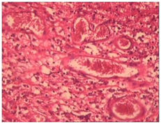
Histopathological examination of hematoxylin and eosin stained soft tissue section revealed. A connective tissue stroma densly infiltrated with chronic inflammatory cells chiefly composed of lymphocytes. (Figure 2) Numerous dilated blood vessels lined by endothelial cells and having erythrocytes inside were seen. (Figure 3) The overlying epithelium was ulcerated (Figure 4).

Based on all the features a final diagnosis of pyogenic granuloma rendered. In 1844, Hullihen described the first case of pyogenic granuloma in English literature.1 In 1897, pyogenic granuloma in man was described as “botryomycosis hominis. in 1904 is credited with giving the current term of “pyogenic granuloma” or “granuloma pyogenicum.2
Differential diagnosis included peripheral giant cell granuloma, peripheral ossifying fibroma, metastatic cancer, hemangioma, pregnancy tumor, conventional granulation tissue hyperplasia, Kaposi’s sarcoma, bacillary angiomatosis and non-Hodgkins lymphoma.3
None.
The author declares no conflict of interest.

©2016 Bajpai, et al. This is an open access article distributed under the terms of the, which permits unrestricted use, distribution, and build upon your work non-commercially.