Advances in
eISSN: 2573-2862


Letter to Editor Volume 2 Issue 1
1SRL Ltd, India
2Department of Pathology, Indira Gandhi Medical College, India
Correspondence: Shailja Puri, SRL Ltd, Shimla, HP, India, Tel 8894363049
Received: November 14, 2016 | Published: January 30, 2017
Citation: Puri S, Kaushik R, Gulati A,et al. Kikuchi’s lymphadenitis: key points in cytology. Adv Cytol Pathol. 2017;2(1):14–15. DOI: 10.15406/acp.2017.02.00009
Kikuchi’s lymphadenitis (KL) is also called histiocytic necrotizing lymphadenitis. KL is a rare but well-defined clinical entity that involves the cervical lymph nodes in young adults. It is a benign condition with a limiting course. The cytology of KL is characteristic but is often missed or misinterpreted as non-specific lymphadenitis. We report a case of KL missed on cytology and diagnosed on biopsy. The fine needle aspiration smears (FNAC) were reviewed and studied carefully. We thus describe characteristic cytological features with which a cytopathologists should be well versed with to avoid missing this non-neoplastic entity.
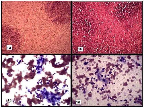
A 25-year-old female presented with painful cervical lymph node enlargement and fever for 15 days. Hemoglobin was 11.5 gm % and fine FNAC was performed. The smears were moderately cellular showing mature lymphocytes and few histiocytes. A cytological diagnosis of non-specific lymphadenitis was given. The patient was put on antibiotics for 15 days after which fever subsided but the lymph node persisted. Thus, a lymph node excision was done. The lymph node measured 2.5 x 2.0 cm. Microscopic examination showed well circumscribed, paracortical necrotizing lesions. The necrotizing lesions showed sheets of mononuclear cells along with abundant karyorrhectic debris and fibrin deposits (Figure 1a & 1b). A histopathological diagnosis of KL was given. FNA smears were reviewed and studied carefully. History revealed a negative Rheumatoid factor. The cellularity of the smears was low, however; histiocytes were evident with apoptotic bodies and peripherally placed eccentric nuclei (Figure 1c & 1d). The histiocytes had a monocytoid appearance. Background showed karyorrhectic debris. Thus, cytologically KL shows a bland population of histiocytes with eccentric nuclei and karyorrhectic debris in the background.

KL was reported in Japan in 1972. Typically KL presents as localized cervical lymphadenopathy in young females.1,2 KL is associated with fever and leucopenia in about 50% of patients.3 The exact etiology of KL is not known but infectious organisms like Epstein Barr virus, Human Herpes virus 6, B-19 Parvovirus, Cytomegalovirus, Human T-cell leukemia virus, para-influenza virus, Brucella and Yersinia have been proposed without any definitive proof.4 KL has been reported in association with autoimmune diseases, thus, raising the possibility of immune mediated mechanism in its pathogenesis. Clinically, fever with cervical lymphadenopathy can be seen in various infections, like tuberculosis, toxoplasmosis, cat-scratch disease, syphilis, HIV; autoimmune conditions and malignancies like lymphoma. FNAC has an important role in distinguishing KL from other causes of lymphadenopathy.
As a rule granulomas, neutrophils, eosinophils and plasma cells are absent in KL. Histiocytes with eccentric crescent shaped non-descript nucleus having monocytoid appearance is a consistent feature of KL. The histiocytes often contain phagocytosed karyorrhectic debris. These histiocytes can be mistaken for tingible- body macrophages found in reactive lymph node.5
The karyorrhectic debris in aspiration smears also raise the possibility of malignancy, however, absence of atypical cells rule out the possibility of malignancy. Thus, an awareness of cytological features of KL among cytopathologists in appropriate clinical context can help in making a precise diagnosis and avoid unnecessary biopsies and surgical excision.
None.
The author declares no conflict of interest.

©2017 Puri, et al. This is an open access article distributed under the terms of the, which permits unrestricted use, distribution, and build upon your work non-commercially.