Advances in
eISSN: 2573-2862


Case Report Volume 1 Issue 1
1Department of Cytology and Gynecological Pathology, Postgraduate Institute of Medical Education and research, India
2Department of Radio diagnosis, Postgraduate Institute of Medical Education and Research, India
Correspondence: Nalini Gupta, Department of Cytology and Gynecological Pathology, Postgraduate Institute of Medical Education and research, Chandigarh, India, Tel 91-172-2755114, Fax 91-172-2744401
Received: November 11, 2016 | Published: December 29, 2016
Citation: Gupta N, Khariwa A, Lal A,et al. Detection of metastatic fibro-lamellar type of hepato cellular carcinoma in ascitic fluid cytology in a liquid-based preparation. Adv Cytol Pathol. 2016;1(1):15–17. DOI: 10.15406/acp.2016.01.00004
Hepato cellular carcinoma (HCC) of fibro lamellar type is an uncommon primary hepatic carcinoma. They occur most commonly in young adults. Their etiology is not clear till date, as they are not associated with chronic liver disease. Fibro lamellar HCC is not an indolent tumor, but has a better prognosis than conventional HCC. We present an interesting case of metastatic fibro lamellar HCC detected in ascitic fluid prepared by liquid-based cytology (LBC). The diagnosis was confirmed by ultrasound-guided fine needle aspiration (US-FNA) of retroperitoneal lymph node. We report a rare case of fibro lamellar HCC detected on exfoliative cytology, which was confirmed by FNAC and immunochemistry (IHC).
Keywords: ascitic fluid cytology, fibro-lamellar hepato cellular carcinoma, fine needle aspiration, immunocytochemistry, liquid based cytology
HCC, hepato cellular carcinoma; US-FNA, ultrasound-guided fine needle aspiration; IHC, immunochemistry; FL-HCC, fibro-lamellar hepato cellular carcinoma; FNAC, fine needle Aspiration cytology; AFP, alpha feto-protein; Hbsag, hepatitis b surface antigen; HCV, hepatitis c virus; MRI, magnetic resonance imaging; IVC, inferior vena cava; MGG, may grünwald- giemsa; H & E, hematoxylin & eosin; CK7, cytokeratin 7
Fibro-lamellar hepato cellular carcinoma (FL-HCC) is a variant of hepato cellular carcinoma (HCC), which accounts for less than 1% of all primary liver cancer.1 It is distinguished from conventional HCC by its distinctive clinicopathological features. Edmondson et al described it as “eosinophilic hepato cellular carcinoma with lamellar fibrosis” in 1956.2 FL-HCC usually occurs at a younger age than conventional HCC.1-3 Conventional HCC is most commonly associated with cirrhosis and viral hepatitis, in contrast, most of the patients of FL-HCC do not have a history of an underlying hepatic disease.1-3 FL-HCC is usually do not present initially with metastases and has a favorable prognosis than conventional HCC.2
Cyto morphological features of FL-HCC on fine needle aspiration cytology (FNAC) have been described earlier.4,5 Detection of this uncommon variant of HCC on exfoliative cytology poses diagnostic challenges to cyto pathologists, especially when there is no clinical suspicion or clinical details are not known at the time of reporting. We present a unique case of metastatic FL-HCC detected on ascitic fluid cytology and ultrasound-guided fine needle aspiration (US-FNA) of retroperitoneal lymph node. We report a rare case of FL-HCC detected on exfoliative cytology.
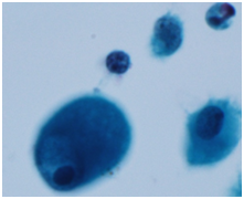
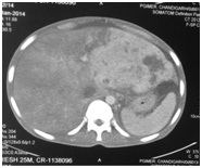
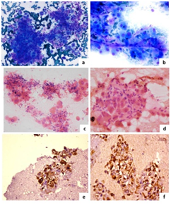
A 32-year-old male presented with loss of appetite and weight with low grade fever since 11 months, pain abdominal since 4 months and abdominal distension since one month. Patient also had hepato splenomegaly and inguinal lymphadenopathy on ultrasonography. Serum alpha feto-protein (AFP) level and HBs Ag antigen levels and anti-HCV were normal. The results of liver function tests were not available at the time of ascitic fluid cytology. Ascites fluid was sent for cytological examination to the Cyto pathology laboratory. The sample was processed by SurePathTM liquid-based cytology (LBC) technique. The smears showed predominantly a few scattered large malignant cells. These cells were polygonal in shape and showed moderate nuclear pleomorphism, round to oval large nuclei, prominent multiple nucleoli, abundant eosinophilic granular cytoplasm (Figure 1). Ascites fluid was reported as positive for malignant cells with suggestion of HCC. CT scan showed an enhancing mass in left lobe of liver with IVC showing hypo dense contents suggestive of thrombus (Figure 2). Ultrasound showed multiple retroperitoneal lymph nodes varying in size from 1 to 2.5 cm in diameter. Liver lesion was not very well-demarcated on ultrasonography. USG guided FNA from retroperitoneal lymph node was performed. Air-dried, MGG-stained (May Grünwald- Giemsa) smears and alcohol-fixed H & E (Hematoxylin & Eosin) stained smears were prepared. FNAC smears were highly cellular smear and showed many individual cells and occasional aggregates of tumor tissue. Individual cells were large with low nuclear to cytoplasmic ratio, had abundant granular and eosinophilic cytoplasm with focal cytoplasmic vacuolations. Tumor cells had round nuclei, coarse chromatin, prominent nucleoli and tumor cells were seen associated with many fibro-collagenous tissue fragments (Figure 3a & 3d). Immunochemistry was performed on cell block, which was prepared from FNA aspirate. The tumor cells were strongly positive for Hep Par 1 and cytokeratin 7 (CK7) (Figure 3e & 3f). Considering the age of the patient [32 years], normal AFP levels, MRI findings, presence of malignant cells with hepatocytic appearance in ascitic fluid cytology and FNAC findings with IHC from retroperitoneal lymph node, a final diagnosis of metastatic FL-HCC was rendered on the USG -FNA sample and ascitic fluid cytology.


FL-HCC is a rare morphological variant of conventional HCC which is mostly not associated with cirrhosis or other chronic liver diseases, and it has a favourable prognosis.2 HCC usually is diagnosed on radiology and /or on hepatic FNAC or core biopsy. Rarely, it may present with metastases. We report a rare case of FL-HCC detected on ascitic fluid cytology prepared by SurePathTM LBC. LBC leads to cellular concentration and therefore, false negative rates are reduced remarkably. Few previous studies have described cyto morphological features of FL-HCC similar to the present case. FNAC smears in case of FL-HCC are usually variably cellular and show predominantly dispersed population of large- sized tumor cells. These cells usually have abundant eosinophilic to granular cytoplasm, centrally placed nuclei, vesicular chromatin pattern, very conspicuous nucleoli and low nuclear to cytoplasmic ratios. Cytoplasmic “pale bodies” represent non-specific cytoplasmic invaginations.6 Intra cytoplasmic hyaline globules and bile can also be present.5 Bland, spindle shaped cells arranged in parallel bands may also be noted on cytology smears, representing collagen fibers and fibroblasts, which are better appreciated on histologic sections. The trabecular arrangement with transversing capillary channels of conventional HCC is not usually seen.4-6 Sometimes, FL-HCC may be difficult to differentiate from conventional HCC. As compared to conventional HCC, the smears in the present case did not reveal the “basketing” pattern” or traversing capillaries that are commonly seen in conventional HCC. Because of characteristic cyto- histological features of FL-HCC’s, this tumor can be readily diagnosed even in the metastatic site. Metastatic FL-HCC has previously been reported in cervical and mediastinal lymph nodes.7,8 There are a few reports in the English literature where a diagnosis of FL-HCC is rendered at uncommon sites. Kunz et al.9 reported a case with tumor involving intra hepatic bile ducts and on biliary brushings a diagnosis was suspected, however, FNAC demonstrated the features of FL-HCC.9 Use of FNAC has rarely been reported in the diagnosis of metastatic FL-HCC.7,8 Detection of F-HCC in effusion sample has been only described rarely. 10In the present case, we prepared cell block and performed IHC which was helpful to clinch the diagnosis. On IHC, the tumor cells of FL-HCC showed positivity for Hep Par 1 similar to conventional HCC but CK7 also showed strong positivity. IHC for Hep Par 1 and CK7 helped to determine the hepatic origin of the tumor, however the same cannot differentiate various variants of HCC. We report detection of fibro lamellar type of hepato cellular carcinoma in ascitic fluid cytology and the diagnosis was confirmed by US-FNA from retroperitoneal lymph node along with IHC on cell block. This case highlights role of cyto pathologist in diagnosing such uncommon tumors at unusual sites.
None.
The author declares no conflict of interest.

©2016 Gupta, et al. This is an open access article distributed under the terms of the, which permits unrestricted use, distribution, and build upon your work non-commercially.