Advances in
eISSN: 2573-2862


Case Report Volume 1 Issue 1
1Department of Pathology, East Tennessee State University, USA
2Pathology and Laboratory Medicine Service, JH Quillen Veterans Affairs Medical Center, USA
Correspondence: Sarah S Kassaby MD, Department of Pathology, East Tennessee State University, PO Box: 70568, Johnson City, TN 37614, USA
Received: September 23, 2016 | Published: December 22, 2016
Citation: Kassaby SS, Azam M, Shurbaji MS. Cellular angiofibroma of the inguinoscrotal region with cytologic atypia and abundant adipose tissue simulating liposarcoma: a case report. Adv Cytol Pathol. 2016;1(1):10–13. DOI: 10.15406/acp.2016.01.00003
Cellular angiofibroma (CAF) is a rare benign mesenchymal neoplasm of the vulvovaginal or inguinoscrotal regions. We report a case of CAF with abundant intralesional fat and focal cytological atypia that raised the possibility of liposarcoma. We review the literature and discuss the significance of these findings and the differential diagnosis of CAF.
Keywords: angiofibroma, differential diagnosis, sarcoma, spermatic cord
CAF, cellular angiofibroma; WDL, well-differentiated liposarcoma; SMA, smooth muscle actin; ER, estrogen receptor; PR, progesterone receptor
Cellular angiofibroma is a benign soft tissue neoplasm that occurs almost exclusively in the vulvovaginal region of women or in the inguinal–scrotal region of men. CAF is characterized by bland spindle cells and prominent small to medium-sized vessels with mural hyalinization. Rarely, cellular angiofibromas have been reported to show atypical or sarcomatous features, including foci resembling undifferentiated pleomorphic sarcoma, and pleomorphic liposarcoma.1,2
A 60 year-old man presented with a one month history of a non-tender, painless, and growing lesion in the inguinoscrotal area that progressively became painful before he presented to medical attention. On clinical examination, a trans-illuminating scrotal mass is found in the scrotum that does not appear to be attached to the epididymis or the testicle. A scrotal ultrasound revealed a solid, heterogeneous and hyper vascular 2.5 cm extra testicular mass in the left lower testicular pole. A scrotal exploration was undertaken with excision of the mass. Grossly, the lesion measured 3 x 2 x 1.5 cm, was gray-tan, firm, well circumscribed, and attached to the spermatic cord. Cut surface of the mass was pink-tan and firm to solid in consistency.
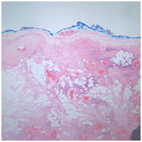
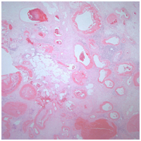
Microscopically, the lesion was well circumscribed (Figure 1) and there were predominately spindle-shaped cells in an edematous and lightly fibrous stroma interspersed with chronic inflammatory cells consisting of lymphocytes with some plasma cells and occasional mast cells. There were numerous thick-walled blood vessels, many with wall hyalinization (Figures 2 & 3). These findings are typical of cellular angiofibroma, but there were also scattered cells with cytological atypia and abundant intralesional adipose tissue with focal fat necrosis (Figures 1 & 4). These additional findings raised concern for a well-differentiated liposarcoma, and the case was referred to the Joint Pathology Center, the reference laboratory for Veterans Affairs medical Centers, for consultation. The consultant performed immunohistochemical stains for CD34, desmin, and estrogen receptor, which were positive, and MDM-2, which was negative. This immuno profile, along with the light microscopic appearance supported the diagnosis of cellular angiofibroma with focal cytologic atypia over well-differentiated liposarcoma.


Cellular angiofibroma (CAF) is an uncommon benign mesenchymal neoplasm of the genital tract with a 2 prominent spindle cell component and very prominent stromal vasculatures. Adipose tissue was present in 24% of cases from one of the largest published series. Adipose tissue was usually located in the periphery of the lesion as small clusters with few cases showing large nests or lobules comprising 30-50% of the tumor area.1 CAF tends to occur in the inguinoscrotal region in males and vulvovaginal region in females. CAF occurs in men and women in equal proportions. It tends to occur in the fifth decade in women and the seventh decade in men.1
In addition to the well-differentiated liposarcoma (WDL) which was included in the differential diagnosis because of some of unusual features of this lesion, the entities that are typically considered in the differential diagnosis of CAF are listed in (Table 1), along with the most useful histologic features that help in differentiating them. Iwasa & Fletcher1 reported immunohistochemical studies on 48 of their study cases. The tumor cells showed multifocal to strong and diffuse positivity for CD34 in 29 cases (60%) (11/24, 46% for female and 18/24, 75% for male cases). Immuno reactivity for smooth muscle actin (SMA) was present in 10 tumors (21%) and for desmin in 4 tumors (8%). The tumor cells were consistently negative for S-100 protein in all cases. Immuno reactivity for estrogen receptor (ER) and progesterone receptor (PR) was examined in 20 randomly selected cases (10 female and 10 male cases). Strong nuclear positivity for ER was present in 7 tumors (35%) (5/10, 50% for female and 2/10, 20% for male cases) and for PR in 11 tumors (55%) (9/10, 90% for female and 2/10, 20% for male cases). (Table 2) summarizes the immunohistochemical profile of the lesions considered in the differential diagnosis of CAF, based on published literature. There is limited genetic data suggesting that CAF appear to be closely related to spindle cell lipoma and mammary-type myofibroblastoma.3,4
Differential Diagnoses of CAF |
Gender |
Distinguishing Histologic Features |
Most Common Site |
Aggressive Angiomyxoma |
F:M=9:1 |
Poorly circumscribed with infiltrative borders |
Genital. Perineal and pelvic region |
Angiomyofibroblastoma |
F:M=20:1 |
Prominent ectatic vessels surrounded by eosinophilic epithelioid cells, usually in vulva |
Genital area |
Spindle Cell Lipoma |
F:M=1:20 |
Mature adipocytes and bland spindle cells. Ropy, loosely arranged collagen |
Posterior neck, upper back and shoulder |
Solitary Fibrous Tumor |
F=M |
Benign tumor of spindle cells separated by thick bands of collagen with hyalinization. Large branching (staghorn) vessels |
Anywhere in the body, especially body cavity sites, including pleura, peritoneum, and meninges |
Neurofibroma |
F=M |
Non-encapsulated. Spindle cells with wavy or comma-shaped nuclei in a wire-like collagenous stroma. Intralesional nerve fibers |
Cutaneous, and anywhere in the body, from the peripheral nervous system |
Table 1 Entities that are typically considered in the differential diagnosis of cellular angiofibroma
Vimentin |
CD34 |
SMA |
Desmin |
ER |
PR |
S100 |
|
Cellular Angiofibroma |
+ |
+ |
+/- |
+/- |
+/- |
+/- |
- |
Aggressive |
+ |
+ |
+/- |
+/- |
+/- |
+/- |
- |
Angiomyofibroblastoma |
+ |
+/- |
+ |
+ |
+ |
+ |
- |
Spindle Cell Lipoma |
No data |
+ |
No data |
- |
No data |
No data |
+(in Adipocytes) |
Solitary Fibrous Tumor |
No data |
+ |
- |
- |
No data |
No data |
- |
Neurofibroma |
+ |
+ |
No data |
No data |
No data |
No data |
+ |
Well Differentiated Liposarcoma |
+ |
+ |
- |
- |
No data |
No data |
Table 2 Immunohistochemical profile of cellular angiofibroma and entities considered in its differential diagnosis
Atypical cellular features and sarcomatous transformation of CAF have been reported.2 CAF with atypical (bizarre) cells shows scattered, noncontiguous foci of severely atypical cells within conventional CAF. The sarcomatous transformation is characterized by abrupt transition to a discrete sarcomatous component that may form a solitary nodule. This sarcomatous component can resemble well-differentiate liposarcoma/ atypical lipomatous tumor, pleomorphic liposarcoma, or pleomorphic sarcoma NOS.2,5 Chen & Fletcher,2 reviewed 154 usual cellular angiofibromas identified between 1993 and 2009 from their consultation files and identified 13 cases of CAF with atypia or sarcomatous change, 12 patients were female and one was male suggesting that atypia and sarcomatous changes are more common in vulvar lesions. Nonetheless, based on limited clinical follow-up data, these changes did not seem to predispose to recurrence or metastasis.2 In that study, three cases of CAF contained areas with features indistinguishable from well-differentiated liposarcoma/atypical lipomatous tumor, but these were negative for MDM2 and CDK4 by immunohistochemistry, suggesting that these two genes are not the underlying alteration when this lesion develops in association with CAF.2 Table 3 summarizes the cases of CAF we identified in our literature search and highlights those that reported atypia.
Case Study/Report |
Number of Cases |
Cases Showing Atypia or Sarcomatous Transformation |
Iwasa et al.1 |
51 (26 Female, 25 Male) |
Mild cytologic atypia characterized by slight nuclear enlargement and hyperchromasia was present in 5 tumors (all males), all of which were associated with stromal edema and a prominent chronic inflammatory cell infiltrate, suggesting that this represented reactive/degenerative atypia. One case (female) showed a focus of a microscopic intralesional nodule of pleomorphic liposarcoma |
Chen et al.2 |
154 (13 cases with atypia described in detail. 12 female, 1 male) |
13 total cases with atypia or sarcomatous transformation. 4 cases of CAF with atypia 9 cases showed morphologic features of sarcomatous transformation including pleomorphic liposarcoma (2 cases), and atypical lipomatous tumor/well-differentiated liposarcoma (3 cases) |
Hameed et al.3 |
1 (male) |
No |
Maggiani et a. 4 |
2 (1 male, 1 female) |
No |
Dikaiakos et al. 7 |
1 (male) |
No |
Val Bernal et al.8 |
1 (male |
CAF showed atypical (bizarre) cells |
Canales et al9 |
2 (males) |
No |
Ptaszynski et al.10 |
1 (male) |
CAF showed atypical cells |
Table 3 Summary of cases of cellular angiofibroma reported in the literature with focus on those showing cytological atypia
Although rare, well-differentiated liposarcoma of the spermatic cord is the most common sarcoma of the paratesticular region in adults.6 The lesion is characterized by scattered, bizarre cells with irregular, hyper chromatic nuclei present in the fibrous stroma or among the adipocytes, or both. These atypical cells may be numerous, or rare and difficult to identify. Typically, adipose tissue predominates (lipoma-like variant), but the adipocytes show more variability in size than mature fat. Lipoblasts may not be seen and are not required for diagnosis. The sclerosing variant, more common in the retro peritoneum, contains predominantly atypical spindle cells in a fine fibrillary collagenous stroma and only scant adipose tissue. Mitoses are rare. Immunohistochemical staining demonstrates nuclear staining for MDM2 (95% of cases) and CDK4 (78% of cases).5,6
In conclusion, the case we present had morphology and immunohistochemical profile consistent with CAF. There were unusual features that included abundant intralesional adipose tissue, in addition to the expected adipose tissue at the periphery. There were also atypical cells that were scattered within the lesion. These atypical cells did not form a discrete nodule, therefore, this lesion is best regarded as cellular angiofibroma7-10 with focal atypia. Iwasa & Fletcher1 suggested that the atypia may be reactive, since most of the lesions, as in our case, had a prominent chronic inflammatory infiltrate. Complete local excision is the optimal treatment for CAF with atypical cells as the lesion shows no tendency to recur based on the current literature. Pathologists should be aware of the morphological variation of CAF and its differentials to avoid misdiagnosis and potential overtreatment.
None.
The author declares no conflict of interest.

©2016 Kassaby, et al. This is an open access article distributed under the terms of the, which permits unrestricted use, distribution, and build upon your work non-commercially.