eISSN: 2378-3176


Case Report Volume 11 Issue 1
Department of urology, University Sidi Mohammed Ben Abdellah, Morocco
Correspondence: Rhyan Ouaddane Alami, Medicine doctor, Department of urology, Hassan II University Hospital, University Sidi Mohammed Ben Abdellah, Fez, Morocco, Tel +212669833956
Received: February 28, 2023 | Published: March 23, 2023
Citation: Alami RO, Mhammedi NA, Ahsaini M, et al. Rare bird spotted: renal clear cell carcinoma in a pancake kidney case report. Urol Nephrol Open Access J. 2023;11(1):20-22. DOI: 10.15406/unoaj.2023.11.00323
The discovery of a tumor process on a Pancake kidney allows the authors to review both the particularities of this association and the diagnostic and therapeutic difficulties. The diagnosis of Pancake kidney may be difficult, and CT Scan examination is very useful to confirm the renal anomaly, tumor situation, and to plan a surgical strategy by visualizing the vascular pedicles which are highly variable. The surgery of exeresis, adapted to the tumor location, represents the essential therapeutic time.
Keywords: pancake kidney, kidney ectopia, renal carcinoma, renal fusion
Pancake kidney (PK) is one of the rarest types of renal anomaly with complete fusion of the superior, mild and inferior poles of both kidneys in the pelvic cavity. Each kidney has its own excretory system with two ureters that do not cross the midline. In the asymptomatic cases, a conservative approach should be performed. Surgical management may be needed when urological problems occur.1 Cancer is a relatively rare entity in this type of malformation with only one pediatric rhabdomyosarcoma having been reported so far in a pancake kidney.2
We report the case of a young patient diagnosed with a renal carcinoma in a Pancake Kidney who underwent bilateral nephrectomy.
A 27-year-old male presented to our service with a 2 year history of intermittent abdominal pain and a palpable abdominal mass. Abdominal ultra-sound revealed a left-sided renal mass. The patient had no fevers, weight loss, vomiting, constipation, appetite change, or change in activity. His medical history demonstrated no previous medical, urologic, or surgical problems. Family history was also unremarkable.
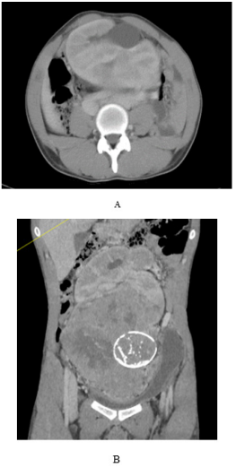
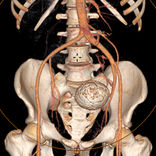
On physical examination, a nontender, firm mass was easily palpable in the left lower quadrant extending to the pelvic brim. Computed tomography Scan of the chest, abdomen, and pelvis revealed a pancake kidney with a large mass in the left moiety, measuring 150X110X200 mm. The tumor extended into the isthmus and appeared to abut or extend into the inferior right moiety, at the site of fusion of both moieties, just below the aortic bifurcation, with a macrocalcification (Figure 1). Bilateral renal arteries were arising from the ipsilateral external iliac arteries (Figure 2) with an accessory renal artery on the right side from the aorta. no metastasis was found.

Figure 1 Contrast-enhanced tomographic scan showing pancake kidney with a tumor at the lower pole with macrocalcification.

Figure 2 Scannographic reconstruction showing the vascularization of the kidney mainly on the iliac axes.
By median laparotomy approach, after location and dissection of the right renal pedicle, an attempt to perform a partial left nephrectomy was unsuccessful, due to the large volume of tumor, and the occurrence of hemodynamic instability, and in front of the commitment of the patient's vital prognosis, a bilateral total nephrectomy was performed with removal of the tumor mass (Figure 3). Histopathology revealed clear cell carcinoma with negative surgical margins. The patient was doing well without recurrence at 6 months, on hemodialysis waiting for renal transplantation.
Pancake Kidney (PK) is one of the rarest types of renal ectopia and fusion anomaly and is more common in men than women.3 Looney and Dodd were the first to define and describe this condition.4 PK is characterized by a large and lobulated renal mass consisting of two fused lateral lobes without an intervening septum located in the pelvic cavity. Each lobe usually has a separate pelvicalyceal system. The renal pelvis is anteriorly placed and the ureters are usually short and enter the bladder normally.5 PK usually receives the blood supply from two main arteries originating from the abdominal aorta, below the inferior mesenteric artery, or from the common iliac artery. A single or additional artery may be present. Venous drainage is carried out by vena cava or iliac vein. In some rare cases, a single renal vein may be found.6
Patients with renal fusion anomalies have been reported to be at an increased relative risk of developing various primary renal tumors including Wilms tumor7 and renal cell carcinoma.8 We postulate that the aberrant embryonic signaling leading to a renal fusion anomaly may place it at risk for malignancy.
Carcinomas are the most frequent.9 While they represent 90% of all kidney cancers (normal kidney), their percentage in cancers on a PK 4is less important and estimated at 54.3%, it seems that this anatomical malformation does not increase the incidence.10
The treatment of such tumor is multimodal. An aggressive surgical approach is encouraged as complete resection is a significant factor in survival.11 The surgical procedure is often difficult because of the non-systematization of this vascularization, variable in number and origin, as well as by the relations of the isthmus which can, in certain cases, pass behind the great vessels or sometimes between the inferior vena cava and the aorta.1 When the tumor is lateralized, the surgical procedure will consist of a heminephrectomy of the portion of the kidney bearing the tumor. During definitive resection, regional lymph node dissection should be performed. In cases of incomplete resection or positive margins after resection, radiation may be offered to reduce the risk of local relapse.2
Pancake kidneys are unusual anatomic anomalies, the occurrence of cancer in this type of malformation poses particular diagnostic and therapeutic problems. Imaging, and in particular arteriography, represents an important step in the surgical strategy by visualizing the different morphological and especially vascular anomalies. Surgery, adapted to the local conditions of PK, is, as in all kidney cancers, the essential therapeutic step.
None.
The authors declare no conflicts of interest.
None.

©2023 Alami, et al. This is an open access article distributed under the terms of the, which permits unrestricted use, distribution, and build upon your work non-commercially.