eISSN: 2378-3176


Case Report Volume 1 Issue 2
1Department of Urology, St George?s Hospital, United Kingdom
2Department of Urology, St George?s Hospital, United Kingdom
3Department of Urology, Ashford and St Peter?s NHS Foundation Trust, United Kingdom
Correspondence: Meghana Kulkarni, St George's Hospital, Core Surgical Trainee Year One, Flat 16, Bentham House, 5 Falmouth Road, London, SE1 4JY, United Kingdom, Tel 07824 345 307
Received: October 27, 2014 | Published: November 8, 2014
Citation: Kulkarni M, Nair R, Kulkarni R. More than one bun in the oven: implications and concerns when managing the unexpected bladder lesion in pregnancy. Urol Nephrol Open Access J. 2014;1(2):38-40. DOI: 10.15406/unoaj.2014.01.00009
Bladder lesions in pregnancy are rare. Whilst the majority represent benign pathology, concern remains regarding a malignant process in the absence of direct visualization or histopathology. In these circumstances, investigation or intervention may risk miscarriage or premature delivery. We discuss the implications of an incidental bladder lesion identified in a 37year-old pregnant female.
Keywords: urinary bladder, endometriosis, pregnancy
MRI, magnetic resonance imaging; LUTs, lower-urinary tract symptoms
The diagnosis of bladder lesions in pregnancy is a rare occurrence. In the vast majority, causes are benign and pregnancy can be completed prior to investigation and treatment, which may be invasive, require ionizing radiation or surgical management. Nevertheless, when identified, concerns surrounding bladder malignancy remain. In these circumstances direct visualization and transurethral biopsy can be both diagnostic and curative. However, there are risks to both mother and foetus which must be considered. We present a case of an incidental bladder mass in a pregnant female and use it to describe the management of a rare but important clinical conundrum.
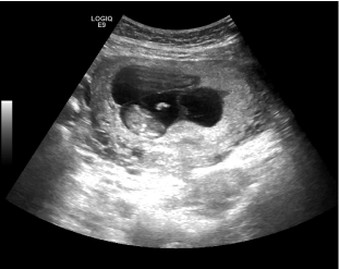
A 37year-old non-smoking primigravid female, underwent routine foetal anomaly Ultrasonography at twenty weeks gestation. This scan confirmed a viable twin pregnancy with normal foetal development, however identified an indeterminate lesion on the posterior bladder wall (Figure 1). The patient did not describe any visible haematuria, nor did she suffer from any lower urinary tract symptoms. She was otherwise in good health with no significant past medical or surgical history.
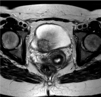
Further characterization of the lesion with pelvic magnetic resonance imaging (MRI) revealed a solid, sub-urothelial two centimetre bladder mass. This was in close relation to the cervix (Figure 2). Concerns between a benign or malignant process led to recommendation of direct visualisation (cystoscopy).


Multidisciplinary involvement between the urological surgeons, obstetricians and the patient allowed for a well-informed rationalized decision. A transurethral en bloc resection of a soft a vascular lesion at the posterior bladder wall under general anaesthetic was performed. The patient was catheterized immediately post-operatively and discharged the following day catheter free. Subsequent histopathological analysis of the resected tissue confirmed endometrial tissue and a diagnosis of urinary tract endometriosis made.
The aims of managing a bladder lesion identified during pregnancy are to determine the origin of the mass and establish if it represents a benign or malignant process. If likely to be malignant, the question remains if investigations or treatment can be safely deferred until after pregnancy is completed. Lesions of the urinary bladder include a broad range of pathologies, from benign masses to primary and secondary malignancies. Common benign bladder lesions include cystitis, nephrogenic adenoma, metaplasia, urachal remnant, endometriosis and polyps. Primary malignancies of the bladder are typically transitional cell carcinoma, squamous cell carcinoma or adenocarcinoma. Secondary malignancies can also occur involving the bladder by direct extension or metastasis. The recognition and distinction of these different entities is challenging but of great clinical importance.1
Endometriosis is a chronic, oestrogen-dependent disease, defined as the presence of functionally-active endometrial tissue in an ectopic location outside of the uterine cavity. Approximately 10% of women of reproductive age suffer from the condition. Urinary-tract endometriosis is rare, with only 1-2% of patients experiencing involvement.2 Lesions are defined as primary or secondary, depending on their onset. Primary lesions are those which are congenitally present in the urinary tract, and tend to present with pain or cyclical haematuria whilst secondary lesions typically occur after birth or pelvic surgery.
Symptoms include storage lower-urinary tract symptoms (LUTs), dysuria and pelvic pain. Cyclical haematuria (menoruria) is present in 20-30% of cases. The presence of endometriosis is often an adverse factor in those who wish to conceive, affecting up to 40% of patients. In general endometriotic lesions regress during pregnancy. Ultrasonography is the initial imaging modality of choice with published data has reporting a sensitivity of between 60-70% in detecting bladder endometriosis, with greater accuracy for lesions is other pelvic sites.3 MRI can aid the further characterisation of lesions, particularly in patients with significant adhesions. There are differing opinions regarding the use of cystoscopy for bladder lesions, as most are located in the submucosal layer and cystoscopic findings may be normal, particularly if performed mid-cycle.
Treatment of the condition is either medical or surgical but must take into account patient-related factors, including age, symptoms and extension of disease, and the wish for future fertility. Medical treatment options are based around the regression of endometrial tissue. This is achieved by gonadotrophin-releasing hormone agonists or antagonists, progestogens or the combined oral contraceptive pill. Surgical treatment is tailored to disease location, and for urinary tract lesions can be endoscopic or radical. Options include trans-urethral resection or ablation or partial or radical cystectomy.4
The primary concern of a clinician faced with any bladder lesion is the question of malignancy. Bladder cancer in pregnancy is rare, with the largest case series of twenty-seven patients reported by Spahn et al.5,6 Three-quarters of cases were superficial and urothelial in origin.6 Painless haematuria remained a key component of presentation in these cases. Haematuria in pregnancy however is a frequent phenomenon which if often correctly attributed to urinary tract infection or confused with per vaginal bleeding. Close visual inspection of urine by the clinician is therefore required by performing urinalysis and culture. If suspicious, urine cytology can be performed as an adjunct; negative cytology however does not exclude an underlying malignancy.7 There are obstetric causes of haematuria that require exclusion in consultation with obstetricians, including placenta praevia, abruption placenta or retained products of conception following an unrecognized miscarriage.8
One must avoid using ionizing radiation in evaluation in pregnancy, and therefore ultrasonography is the initial imaging modality of choice. Ultrasound is usually able to provide accurate anatomical information, with a prospective study confirming a sensitivity and specificity of 91% and 79% respectively for the detection of bladder lesions. The gravid uterus however can sometimes make this investigation challenging to interpret. Magnetic resonance imaging (MRI) is an excellent alternative tool to evaluate the bladder and can give accurate information regarding the transmural spread of bladder lesions. Provided urine cultures are negative, flexible cystoscopy under local anaesthetic is safe and highly effective in evaluating intravesical pathology. The role of procedures such as resection requiring regional or general anaesthesia has also been deemed safe in pregnancy.9 Managing malignancy in pregnant woman is very complex and whilst there are there are no randomised trials available to guide clinicians, the current published data has suggested that cancer rarely comprises pregnancy unless the patient is at a terminal phase of disease. Treatment can therefore usually be deferred until delivery.9
When considering a low-grade urothelial malignancy, most cases are superficial and less than two percent progress to muscle invasive disease.6 In cases where patients are symptomatic (haematuria or recurrent urinary tract infections threatening pregnancy), or if muscle invasive or high-grade disease is suspected (i.e. previous history of high grade non muscle invasive disease, genetic conditions predisposing to bladder cancer or imaging suggestive of transmural disease) endoscopic evaluation and resection should be considered urgent. Here collaboration and involvement of the patient, urologists, obstetricians and midwives is crucial to adequately inform the patient and partner of the risks of lower urinary tract instrumentation under regional or general anaesthesia.
The investigation and treatment of a bladder mass in pregnancy can result in divided opinion, and requires an individualised multidisciplinary approach to patient care. Involvement of the patient, urological surgeons, obstetricians and midwives is considered a key to ensuring expectations and concerns are addressed. The risks of unearthing and treating a potential malignant process versus that of expectantly manage a potential indolent bladder lesion must be weighed against the risk of premature labour or miscarriage.
There are no financial or other conflicts of interest.

©2014 Kulkarni, et al. This is an open access article distributed under the terms of the, which permits unrestricted use, distribution, and build upon your work non-commercially.