MOJ
eISSN: 2475-5494


Case Report Volume 1 Issue 3
Department of Obstetrics & Gynaecology, Universiti Sains Malaysia (USM), Malaysia
Correspondence: Ahmad Shuib Yahaya, Obstetrics & Gynaecology Department, School of Medical Sciences, USM Health Campus, 16150 Kubang Kerian, Kelantan, Malaysia, Tel 60167767974
Received: October 30, 2015 | Published: December 29, 2015
Citation: Yahaya AS, Khairuddin NK. Uterine compression suture; new technique using parenchymal set suture. MOJ Womens Health. 2015;1(3):60-61. DOI: 10.15406/mojwh.2015.01.00014
Over 125,000 women die of postpartum hemorrhage each year. Important cause is uterine atony and traditional management of this condition includes the use of oxytoxics before proceeding to ligation of internal iliac & uterine arteries and even hysterectomy. Since B-Lynch suture introduced in 1997, there had been many reported new techniques and modification of uterine compression sutures.
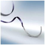
We reported a case of primigravida in term pregnancy, presented with antepartum haemorrhage due to abruptio placenta complicated with fetal distress. Emergency caesarean section was done and complicated with uterine atony, not responsive to pharmacological therapy. We performed uterine compression suture using technique almost similar as B-Lnch, with modification in suture type used. We used a special parenchymal set manufactured by B-Braun (Figure 1), usually used for adaptation and haemostasis of parenchymal organs such as liver, kidney and spleen. Advantages include wide tape-like suture with its outstanding flexibility and softness lead to excellent tissue approximation, easier knotting and prevention of trauma to uterus by sharp edge of normal suture. The long blunt needle allows for safe handling and suture placement.
Keywords: B-lynh, uterine compression sutures
Over 125,000 women die of postpartum hemorrhage each year. Important cause is uterine atony and traditional management of this condition includes the use of oxytoxics before proceeding to ligation of internal iliac & uterine arteries and even hysterectomy. Since B-Lynch suture introduced in 1997,1 there had been many reported new techniques and modification of uterine compression sutures.
We reported a case of primigravida in term pregnancy, presented with antepartum haemorrhage due to abruptio placenta complicated with fetal distress. Emergency caesarean section was done and complicated with uterine atony, not responsive to pharmacological therapy. Intramuscular boluses of Syntometrine were given, followed by 3 boluses of intramuscular Haemabate 250mcg (carboprost). Continuous infusion of Oxytocin 40units in 500ml saline was running.
Uterine compression suture done using parenchymal set suture, an absorbable Safil® violet tape of polyglycolic acid, attached to a round bodied blunt needle. Uterine compression and haemostasis were able to achieved and patient had good outcome without need of further intervention and massive blood transfusion. Total blood loss was 1000ml and patient was maintained haemodynamically by fluid infusion.
Tehnique
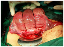
Uterine compression suture using technique almost similar as reported by B-Lynch et al.1 with modification in suture type used. We used a special parenchymal set manufactured by B-Braun (Figure 1), usually used for adaptation and haemostasis of parenchymal organs such as liver, kidney and spleen. Advantages include wide tape-like suture with its outstanding flexibility and softness lead to excellent tissue approximation, easier knotting and prevention of trauma to uterus by sharp edge of normal suture. The long blunt needle allows for safe handling and suture placement (Figure 2).
With the bladder pushed inferiorly, the first stitch is placed approximately 3cm below the uterine Caesarean incision (transverse, lower segment) on maternal right side and threaded through the uterine cavity to emerge anteriorly 3cm above the upper-incision margin. The suture should be placed approximately 4cm from lateral border of the uterus, to avoid large branches of uterine vessels running vertically. The suture now carried over to the fundus of uterus to the posterior side and it should be more or less vertical overlying the uterine fundus. The assistants must compress the uterus while suturing is performed, to ensure proper placement is achieved and maintained. The suture is brought posteriorly downwards and should be pulled until point at the level of uterine incision at the insertion of the uterosacral ligament. The suture is then fed through the posterior wall into the uterine cavity and it should be brought horizontally on the cavity side of the posterior wall to the left margin. The suture is pulled again through the posterior wall, brought over the top of the fundus posteriorly, an then down the anterior left side of the uterus. The needle is now placed in a position symmetrical to that in which it first entered the right side (i.e., 3cm above the upper lip of the incision and 4 cm from the lateral side of the uterus), pushed into the uterine cavity and then again through the lower segment, 3cm below the incision margin.
Uterine incision is now need to be closed in the usual two layers, continuous non-locking technique using an absorbable suture (we use Safil ‘1’ braided suture). The two ends of the compression suture (parenhymal set suture) will be then tied with a double throw knot to maintain tension after the lower segment incision had been closed.


The uterine compression sutures can preserve life and fertility and it has been recommended by various authorities worldwide. The chance of success is high and the procedure can be performed by surgeons with average surgical skills at units with limited resources. Since B-Lynch suture introduced, various other techniques had been described, such as Hayman suture2 and Cho suture.3 The technique being outlined in this report is following B-Lynch steps (although initially described using catgut suture material), but we proposed the parenchymal set suture can also be used in performing Hayman suture (90cm length).
Uterine compression sutures may provide a simple first surgical step to control bleeding when routine pharmacological measures have failed. They have proved to be valuable in the control of massive PPH as an alternative to hysterectomy. We suggest that the technique we described should be tried before more complex interventions are used.
Head, O&G Department, USM, Operation theatre staffs, Hospital USM
The author declares no conflict of interest.

©2015 Yahaya, et al. This is an open access article distributed under the terms of the, which permits unrestricted use, distribution, and build upon your work non-commercially.