MOJ
eISSN: 2641-9300


Case Report Volume 1 Issue 1
1Department of Medicine, New Hope Medical School, Brazil
2Department of Medicine, New Hope Medical School, Brazil
3Department of Cardiology and Semiology, University Center of Jo
4Department of Anesthesiology, Para
5Department of Vascular Surgery, Federal University of Paraiba, Brazil
Correspondence: Paulo Roberto da Silva Lima, Department of Cardiology and Semiology, University Center of Joao Pessoa, Brazil
Received: February 01, 2018 | Published: February 16, 2018
Citation: Leite PHCM, Albuquerque MSC, Silva AKM, et al. Palm surgery in iliac vein occlusion: case report. MOJ Tumor Res. 2018;1(1):31-32. DOI: 10.15406/mojtr.2018.01.00005
Palm surgery was first described in 1958 as a therapeutic option for post-phlebitis syndrome. This procedure aims to relieve the high venous pressure established due to a chronic occlusion of large veins draining from the lower limb, and also to prevent the recurrence of post-thrombotic syndrome. This surgical modality is reserved for those cases resistant to pharmacological treatment for more than 12 months; with a deep femoral vein patent on the affected side; and pressure gradient between the two common femoral veins above 10 mmHg.
Case presentation: A 43-year-old male patient referred with a history of cutaneous lesion in the right thigh, with an anatomic pathology already performed, indicating ulcerated malignant melanoma, in vertical growth phase, showing Clark IV index, Breslow index of 2.5 mm, with angiolymphatic and perineural invasion present. A computed tomography scan of the lower abdomen with contrast was required, showing an expansive process in the right pelvic cavity, measuring 9.0 x 7.0 cm in the largest transverse diameters, with presence of intra-lesional necrotic component. The lesion exerts a compressive effect on the ureter, causing moderate hydronephrosis on the right, and involves the ipsilateral iliac vessels, causing occlusion of the iliac veins on the right. The conduct adopted, due to the nonresectability of the tumor, was the Palm surgery, using an autologous internal saphenous vein graft for correction of blood flow.
Conclusion: The present report becomes relevant, since access to a vein through the tumor endovascular is practically impossible, so it is necessary to perform this procedure to divert the flow and allow drainage of the affected limb. Palm surgery, although old, is still an excellent therapeutic option to avoid more severe presentations of venous obstruction, such as phlegmasia.
Keywords:venous occlusion, palm graft, vascular graft
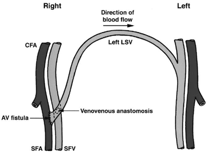
Palm surgery was first described in 1958 as a therapeutic option for the post-phlebitis syndrome. This procedure aims to relieve the high venous pressure established due to a chronic occlusion of large veins draining from the lower limb, and also to prevent the recurrence of post-thrombotic syndrome, being reported in the literature 90% success rates and patency of 83 % after four years. The technique aims to perform a crossed femorofemoral venous bypass,1 where the internal saphenous vein of the contralateral limb is mobilized, supra-pubic, to the affected limb, when it is anastomosed with the common femoral vein on the obstructed side, thus performing the blood flow deviation from the compromised limb to the other limb where drainage of the lower limb has no obstruction in the iliac limbs. This surgical modality is reserved, nowadays, for those cases resistant to pharmacological treatment for more than 12 months; with a deep femoral vein patent on the affected side; and pressure gradient between the two common femoral veins above 10 mmHg and in cases where there is no way to perform an endovascular procedure to clear the impaired iliac vein, such as extrinsic compression.
A 43-year-old male patient with a history of cutaneous lesion in the right thigh, with an anatomopathological examination showing ulcerated malignant melanoma, in a vertical growth phase, showing Clark IV index, Breslow index of 2.5 mm, with angiolymphatic and perineural invasion present, hepatic and bone metastasis in various segments throughout the body. He came to the office with large edema all over his right lower limb (Figure 1). A color Doppler ultrasonography of the right lower limb venous system was performed, which demonstrated extrinsic occlusion in the transition with the right external iliac vein, and a computed tomography scan of the lower abdomen with contrast was performed, evidencing an expansive process in the right pelvic cavity, measuring 9.0 x 7.0 centimeters in the largest transverse diameters, with presence of intra-lesional necrotic component (Figure 2). The lesion exerts a compressive effect on the ureter, causing moderate hydronephrosis on the right, and involves the ipsilateral iliac vessels, causing occlusion of the iliac veins on the right, but without compromising the arterial lumen. The conduct adopted, due to the nonresectability of the tumor and the impossibility of performing venous angioplasty, was the Palm surgery (Figure 3-6), using an autologous left internal saphenous vein graft to correct blood flow draining the right lower limb to the left lower limb and confection of arteriovenous fistula distally to the veinvenous anastomosis to prevent recent venous graft thrombosis. In this patient, the venovenous anastomosis and arteriovenous fistula of the right lower limb were performed in the middle third of the right superficial femoral vein in order to tumor invasion in the area of the right common femoral vein, which prevented the dissection and isolation of the right common femoral vein. The patient evolved with regression of edema in the right lower limb and improvement of the quality of life of the patient. Due to the advanced state of the mestastatic melanoma and general worsening of the patient, he died four months later.2,3

The present report becomes relevant, since access to a vein through the tumor endovascular is practically impossible, so it is necessary to perform this procedure to divert the flow and allow drainage of the affected limb. Palm surgery, although old, is still an excellent therapeutic option to avoid more severe presentations of venous obstruction, such as phlegmasia.
None.
Authors declare there is no conflict of interest in publishing the article.

©2018 Leite, et al. This is an open access article distributed under the terms of the, which permits unrestricted use, distribution, and build upon your work non-commercially.