MOJ
eISSN: 2379-6162


Case Report Volume 2 Issue 2
Department of Surgery, University Hospital Galway, Ireland
Correspondence: Manvydas Varzgalis, Department of Surgery, University Hospital Galway, Newcastle Road Galway, Ireland, Tel +353871623327
Received: March 30, 2015 | Published: April 18, 2015
Citation: Varzgalis M, Halloran ON, Lowery A, et al. Spontaneous rupture of the inferior pancreatoduodenal artery: case report and literature review. MOJ Surg. 2015;2(2):47-49. DOI: 10.15406/mojs.2015.02.00017
Idiopathic spontaneous intraperitoneal haemorrhage is rare cause of non-traumatic intra-abdominal bleeding and carries high mortality risk if untreated. It is quite challenging to make preoperative diagnosis especially if patients present in hemodynamically unstable state. We report an unusual case of idiopathic spontaneous intraperitoneal haemorrhage caused by rupture of inferior pancreatoduodenal artery which was managed using novel approach with resuscitative endovascular balloon occlusion of the aorta.
Keywords: inferior pancreaticoduodenal artery, spontaneous rupture, surgery, embolization, intra-aortic balloon pump, resuscitative endovascular balloon occlusion of the aorta
ISIH, idiopathic spontaneous intraperitoneal haemorrhage; ICU, intensive care unit; WCC, white cell count; Hb, haemoglobin; FAST, focused assessment by sonography in trauma; IABP, intra-aortic balloon pump; REBOA, resuscitative endovascular balloon occlusion of the aorta
Idiopathic spontaneous intraperitoneal haemorrhage (ISIH) represents a rare cause of acute abdominal pain with haemodynamic instability or shock. This life-threatening surgical emergency is historically referred to as “abdominal apoplexy”. Clinical presentations include a variety of symptoms such as abdominal pain, shock and hematochezia, however the pre-operative diagnosis remains challenging and emergency surgical exploration is often mandatory. Every patient with hypotension and abdominal pain without history of trauma should have spontaneous intraperitoneal haemorrhage included in differential diagnosis. We present a case of ISIH caused by spontaneous rupture of the inferior pancreaticoduodenal artery.
A 54 year old woman presented with sudden onset of severe right sided abdominal pain. The pain, which was described as “burning” had started six and half hours previously, initially in the right upper quadrant with subsequent radiation to the back and right iliac fossa. There had been three associated episodes of vomiting. The patient had no past abdominal surgery or significant medical history. At the time of admission, the blood pressure was 98/65 mmHg, and her pulse rate was 98b/min with a temperature of 37°C.
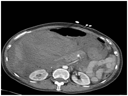
On physical examination there was mild abdominal distension with right sided abdominal tenderness with guarding and present bowel sounds. Blood tests on admission were mildly deranged with white cell count (WCC) of 12.2g/dL neutrophils 10.7%, haemoglobin (Hb) 11.7g/dL, C Reactive Protein<0.6, amylase 28u/L and lactate 0.9mmol/l. Two hours later the patient deteriorated clinically with a fall in BP to 88/50, diaphoresis, Hb dropped to 6.6 g/dL and her lactate rose to 2 mmol/l. The sudden decrease in Hb suggested her deterioration was of haemorrhagic origin. She was transferred to ICU for resuscitation and once she had been stabilised a CT scan was undertaken to determine the source of bleeding. Contrast-enhanced abdominal computed tomography showed a massive intra-abdominal haemorrhage with active extravasations closest to the branches of the superior mesenteric artery especially inferior pancreatic duodenal artery (Figure 1).

It was decided to proceed with an emergency exploratory laparotomy, due to the patient’s transient response to resuscitation. A multidisciplinary team of general and vascular surgeons were involved. Prior to laparotomy, on table CT angiogram was performed using the right CFA approach. A reliant balloon (Medtronic) was placed in the descending aorta through 12 F sheath and was inflated to appropriate diameter to occlude descending thoracic aorta followed by laparotomy using an upper midline incision. A massive intraperitoneal hematoma of 2 litres was evacuated, as there was large retroperitoneal haematoma as well, duodenum was mobilised using the Kocher method. Once the duodenum was mobilised and balloon was deflated the sudden haemorrhage from a tear in the inferior pancreatic duodenal artery was identified at the head of the pancreas at the junction of D2/D3. This vessel was transfixed with 5/0 prolene to achieve haemostasis. A biopsy of the vessel was also sent for histopathology but revealed no specific findings of arteriosclerosis, vasculitis, infection or underlying collagen/aneurismal disease. The patient’s post-operative course was uneventful and she was discharged on the 12th post-operative day.
Idiopathic spontaneous intraperitoneal haemorrhage (ISIH) was first reported by Barber1 and later named “abdominal apoplexy” by Green et al.2 ISIH can occur at any age and is more common in males, the maximum prevalence being at age 50-59.3 There are numerous causes of spontaneous non-traumatic abdominal haemorrhage including aneurysmal disease, pregnancy, vasculitis, arteriosclerosis, malignancy, and inflammatory processes. It is believed that in older patient’s atherosclerosis is the main cause while defects in the media of splanchnic vessels have been cited as a more common cause in younger patients. Rupture of an inferior pancreaticoduodenal artery in the absence of these conditions is an extremely rare cause of ISIH.4
The presentation of ISIH is variable. The most common symptoms include abdominal pain which can be diffuse or localised and distension caused by an expanding haematoma. Bleeding might be intraperitoneal, retroperitoneal or a combination of both.5 Most common causes in the absence of aneurysm are necrotizing arteritis seen with polyarteritis nodosa and rheumatoid arthritis with hypertension, arteriosclerosis being risk factors.6,7 It can occur at any age from 2 to 84 and is more common in males the maximum prevalence at age 50-59.8 It is believed in older patients atherosclerosis is the main cause while in young patients’ defects in the media of splanchnic vessels.4
Hypotension and other signs of hypovolemic shock may become evident at a later stage. This may coincide with a drop in haemoglobin, and as in this case, a non-specific finding of leukocytosis is frequently noted on the patient’s haematological work up. Signs of shock and abdominal wall bruising would relate to rapidity of exsanguination and mostly develop in later stage.
Radiological evaluation includes diagnostic FAST (focused assessment by sonography in trauma) examination followed by surgical intervention in hemodynamically unstable patient. If the patient is stable enough CT scan with oral and i.v. contrast would be best diagnostic tool as we showed it in our case.8 It is not recommended to use the FAST exam in replacement of a CT scan in stable patients. Studies recorded a sensitivity of FAST between 40-99%, where as the CT scan has a documented sensitivity of 92-98%.8,9
The treatment of ISIH centres on appropriate and effective patient resuscitation and definitive control of the bleeding source. Trans-arterial embolization has recently been described as the treatment of choice if resources are available in. If no such resources available exploratory laparotomy and surgical repair should be used.7
The traditional approach to massive intraperitoneal haemorrhage involves clamping aorta at the level of the diaphragm or through the gastrohepatic ligament. This is a technically difficult manoeuvre even in the hands of an experienced surgeon.11 In this case we used a novel minimally invasive vascular approach by endovascular-aortic balloon occlusion. This facilitated safe dissection and mobilisation of the duodenum to reveal the bleeding vessel, while maintaining haemodynamic stability. This was a major advantage in our case, it let kocherise bloodless plain and maintain haemodynamic stability.
There are reports of successfully using temporary intra-aortic balloon pump (IABP) or resuscitative endovascular balloon occlusion of the aorta (REBOA) for patients in haemorrhagic shock as adjunct treatment in the emergency department or in the operation theatre.12 It might be used before laparotomy if the systolic blood pressure remains less than 80mmHg despite adequate resuscitation or during laparotomy if the blood pressure decreases below 60mmHG.13,14 The successful use of IABP/REBOA has also been reported in life-threatening haemorrhagic shock from pelvic fractures.15 This technique requires basic surgical and catheter skills13 and should be considered for incorporation in the surgical training curriculum as it is superior to intra thoracic or intraabdominal aortic clamping.14–17 With evolving technology successful results were shown on fluoroscopy free REBOA on animals.16 Recent study of one centre experience suggested that hemodynamically unstable patients with abdominal solid organ injury could be treated non-operatively with angioembolization and IABP/REBOA.17
Spontaneous non-aneurysmal inferior pancreatoduodenal artery rupture is very rare.18,19 To our knowledge, this is the first report of spontaneous rupture in a non-aneurysmal pancreatoduodenal artery.20,21 The appropriate management principles of the patient would involve the same, initial resuscitation and definite control of the bleeding cause depending on institution capacities although novel techniques of IABP/REBOA and angioembolization are becoming alternative to conventional laparotomy.
None.
The author declares no conflict of interest.

©2015 Varzgalis, et al. This is an open access article distributed under the terms of the, which permits unrestricted use, distribution, and build upon your work non-commercially.