MOJ
eISSN: 2379-6162


Case Report Volume 12 Issue 3
Department of Oncology Surgery, Grupo Hospitalar Conceição, Porto Alegre, Brazil
Correspondence: Francisco Loes, Department of Oncology Surgery, Grupo Hospitalar Conceição, Porto Alegre - RS, Brazil
Received: December 09, 2024 | Published: September 30, 2024
Citation: Loes F, Urzagaste CHV, Filippi LT, et al. Retroperitoneal solitary fibrous tumor: case report. MOJ Surg. 2024;12(3):97-98. DOI: 10.15406/mojs.2024.12.00271
In this, a reported case is presented of a young heathy woman, who incidentally figured out a tumor four months after undergoing a computed tomography scan following a hysterectomy for a benign cause. This huge tumour is very rare and its frequently present in pleural tissues, the main treatment is resection and if the resection is R0 it would have good prognostic. A solitary fibrous tumor is a rare neoplasm of mesenchymal origin that has not been extensively studied, resulting in a limited number of articles on the topic.
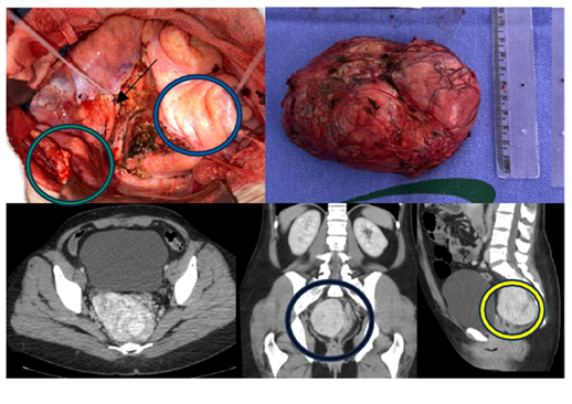
A 45 years old Female, smoker, experiencing depression with a history of previous surgeries: hysterectomy for fibroids and previous dermolipectomy. She started investigation for low back pain, constipation, increased abdominal volume and weight loss of 4kg within 4 months of evolution. Computed tomography (CT) (Figure 1) and MRI showed a voluminous solid expansive lesion located between the rectum and the pre-sacral space, measuring 9.9 X 7.1 X 8.6 CM, slightly to the right of the midline, with no signs of metastasis. During the colonoscopy, there was slight extrinsic compression of the intestinal wall in the middle and lower rectum but no changes were observed in the mucosa. A percutaneous biopsy confirmed the diagnosis of a solitary fibrous tumor.

Figure 1 Tumor syntopy (yellow circle), in relation to the rectosigmoid (blue arrows) and bladder (green circle) and projection into the pelvis (black circle) and right ureter (black arrow).
The patient underwent surgical resection of the lesion via laparotomy, passing a double J catheter preoperatively. She had a good evolution and was discharged on the 5th postoperative day without any complications. The anatomopathological analysis revealed a low-grade mesenchymal neoplasm, measuring 11 cm at its longest axis, with two mitotic figures per field and a capsular surgical margin. Immunohistochemistry (IHC) confirmed the diagnosis of a solitary fibrous tumor (Ki67 at 5%). During the outpatient follow-up 6 months after the resection, there were no signs of recurrence.
CD34 (clone QBEnd 10): Positive
STAT6 (clone EP325): Positive
S100 (polyclonal): Negative
Desmina (clone D33)- Negative
Ki-67 (clone MIB-1): Positive (5%)
CD 99, MIC2 (clone EPR3097Y): Positive
CD31 (clone JC70A): Negative
Conclusion
Immunohistochemical profile of bell-shaped solitary fibrous tumor
The solitary fibrous tumor (SFT) was described for the first time in 1931 by Klemperer and Rabin as a pleural neoplasm.1,2 This tumor is a neoplasm of mesenchymal origin, very rare, corresponds to about 2% of soft tissue tumors and can occur in several syndromes, with pleural cases being the most common.3 There is no gender predilection, but it is more frequently found in individuals aged 50 to 60. Benign forms are characterized by spindle cells with low mitotic activity and low nuclear atypia, while malignant forms exhibit high cellularity, increased nuclear pleomorphism and augmented mitotic activity.4
Diagnosis is suspected through imaging studies and must be confirmed by percutaneous biopsy. The presence of the NAB2-STAT 6 fusion gene in immunohistochemistry brings high sensitivity and specificity for the diagnosis. Surgical resection with free margins is still the standard treatment.2,3 Lymph node metastases are not frequent so lymphadenectomy is not routinely performed.5
This paper presents a case of a solitary fibrous tumor resected with free margins and without recurrence in 6 months. The behavior of these tumors vary based on risk factors: tumor larger than 10cm, higher mitotic index, tumor necrosis, older age at diagnosis, extra-thoracic location.2,3
Given the rarity of this lesion, further studies are necessary to determine the optimal management strategies for solitary fibrous tumors and the potential role of systemic treatments. Due to their low responsiveness to conventional chemotherapy and radiotherapy, research studies suggest the use of anti-angiogenic therapy in metastatic cases.6,7
None.
The authors declare that there are no conflicts of interest.

©2024 Loes, et al. This is an open access article distributed under the terms of the, which permits unrestricted use, distribution, and build upon your work non-commercially.