MOJ
eISSN: 2379-6162


Case Report Volume 5 Issue 2
1Trauma and Orthopedics Department, Hospital Vila Franca de Xira, Portugal
2Trauma and Orthopedics Department, Hospital Dona Estefânia, Portugal
Correspondence: Pedro Campos, Trauma and Orthopedics Department of Hospital Vila Franca de Xira, Estrada Carlos Lima Costa, N, Tel 003-519-146-333-98
Received: August 10, 2017 | Published: November 9, 2017
Citation: Campos P, Rosa B, Barros A, et al. Nora’s lesion: a rare tumor in the pediatric population. MOJ Surg. 2017;5(1):171-173. DOI: 10.15406/mojs.2017.05.00103
Nora’s lesion is a rare benign osteocartilaginous lesion characterized by a mixture of immature bone cells, atypical and bizarre cartilage cells and bland spindle cells. Its pathogenesis remains controversial and its associated signs and symptoms are variable. It is important to identify its clinical, radiological and histopathological characteristics to make the differential diagnosis with other benign or malignant diseases. There are important considerations regarding its diagnosis, which will guide treatment and follow-up. The primary treatment for Nora’s lesion is surgical excision. Despite its high rate of local recurrence, the occurrence of metastasis was not reported. We present the case of an adolescent with the diagnosis of Nora’s lesion located on the terminal phalanx of the first toe. Our aim is to alert for the existence of this condition in younger ages and in atypical locations and for the need to establish the differential diagnosis with other pathologies, providing the patient the most appropriate treatment.
Keywords: nora’s lesion, bizarre periosteal, osteochondromatous, proliferation, terminal toe phalanx, recurrence, neoplastic process
Nora’s lesion or bizarre periosteal osteochondromatous proliferation, first described by Nora in 1983,1 is a very rare condition with only about 160 cases being described in the literature since then.2,3 It is more common in adults, in their third to fourth decade1,2,4 and occurs mostly on the proximal and middle phalanges, metacarpal and metatarsal bones.5 The etiology of this lesion remains unknown. Some authors hypothesized that traumatic events could be on the origin of it.6 However, actually there is no evidence of a relationship between trauma and bizarre periosteal osteochondromatous proliferation. Radiologically, it appears as a calcified mass attached to the bone cortex1 and can mimic an exostosis, an osteochondroma or a malignant condition.7
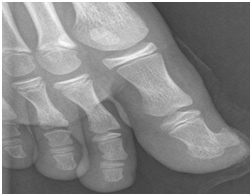
A 12-year-old boy presented at our Institution with right toe pain and swelling with six months of evolution. The patient also presented a progressive increase in pressure sensation. He had no history of trauma prior to the emergence of the symptoms. On physical examination, the terminal extremity of the second toe was swollen, without any other signs such as heat or erythema. The nail plate was elevated, hyperkeratotic and thickened. The toe mobility, sensibility and vascularity were normal. Radiographs showed a heavily calcified mass on the terminal phalanx of the first toe, which was attached to the underlying cortex (Figure 1). There were no signs of periosteal reaction and no alteration of the underlying bone to which the lesion was attached. In this sense, a presumptive diagnosis of subungual exostosis was made and we decided to perform its surgical resection. We decided to perform a subungual approach, lifting the nail plate, which allowed a dorsal view of the terminal toe phalanx. An “en-bloc” resection of the lesion and a decortication of the underlying bone were performed. We have also removed the portion of the nail which presented alterations.

The dimensions of resected segment (excluding the nail) were 1,3 x 1,8 x 1,1 cm and the lesion was well circumscribed and presented a narrow base (Figure 2). The histopathology revealed areas of hypercellular cartilage, which presented a bluish color, large chondrocytes and endochondral ossification. Moreover, bony trabeculae presented a rim of osteoblasts and the intertrabecular spaces were filled with fibroblastic tissue. There were no signs of malignancy. These findings were consistent with the diagnosis of Nora’s lesion (Figure 3). The postoperative radiography did not show any signs of lesion. The patient did not receive any other therapy. At this moment, one year after surgery, the patient has no symptoms or signs of lesion recurrence.
Nora’s lesion is a rare condition and there are some doubts regarding its etiology and evolution. It is described as a distinct entity containing atypical and bizarre cartilage that often undergoes a characteristic irregular ossification5. It occurs mainly in adults and its occurrence in children and adolescents was only rarely described in the literature.8 However, when occurring in younger ages, some of the lesions can grow in their size, causing angular deformities.9 According to cases already described in the literature, Nora’s lesion is more frequent on feet and hands, being the affection of the terminal phalanx of the toes very unusual.1 We describe a case of an adolescent who presented with an osteocartilaginous lesion on the terminal phalanx of the first toe below the nail plate. Its uncommon location could reduce the index of suspicion for the diagnosis of Nora´s lesion. Its gross appearance was similar to that of a small osteochondroma or a subungual exostosis. At first, according to physical examination and radiological findings, we admitted the diagnosis of subungual exostosis. However the histopathological examination revealed findings consistent with bizarre periosteal osteochondromatous proliferation or Nora’s lesion.
In fact, there are some conditions which are more frequent and present similarly to Nora´s lesion, so we must be aware and know how to make this differential diagnosis. In this regard, some radiological exams can be valuable. On the radiography, Nora’s lesion presents as a calcification or as an ossification which seems to arise directly from the cortical surface of the bone without disturbing its native architecture.1 Other exams, such as Computed Tomography (CT-Scan) or Magnetic Resonance (MRI), can give more detailed information in order to plan a correct therapeutic approach.10 On MRI, Nora’s lesion presents with bone cortex involvement without medullary involvement, periosteal reaction or soft tissue swelling.11 However, the definitive diagnosis is given by the histopathological examination. Moreover, this will inform about its behavior and prognosis. Histopathologically the lesion had three components: fibroblastic spindle cells, bluish cartilage with mild cellular atypia and irregular bony trabeculae, all presented in intermixed islands without any specific pattern.8,12 As described in the literature, these findings are typical, however some cases with histological variability were described.7 Subungeal exostosis and osteochondroma frequently can mimic Nora´s lesion. Subungual exostosis occurs mostly on the terminal phalanx of the great toe. It may be preceded by a history of trauma and usually presents as a painful nodular growth or as an externally visible localized lesion that erodes the overlying tissue. Plain radiography normally shows an outgrowth with trabeculated pattern of cancellous bone with or without defined cortex. Histopathologically, it also shares some features with Nora’s lesion.3,5 Osteochondroma is another benign lesion common on toes. Unlike Nora´s lesion, radiologically, the typical osteochondroma presents medullary continuity between the lesion and the native bone, and histopathologically, an organized cap with hyaline cartilage is observed.13 Other differential diagnosis include enchondromas, glomus tumour, myositis ossificans, florid reactive periostitis and heterotopic chondro-ossification.3,14–16 Moreover, Nora’s lesion must be differentiated from malignant tumours such as chondrosarcoma, osteosarcoma or osteochondroma with malignant transformation.13 In fact, the lesion’s histopathology, with marked proliferative activity, irregular bony-cartilaginous interfaces and enlarged bizarre and binucleat chondrocytes, could mimic a chondrosarcoma.
A surgical “En-bloc” resection is the recommended treatment for Nora’s lesion2 and that was our procedure of choice in this reported case. Resection of its capsule and decortication of the underlying cortical bone is reported as fundamental to reduce recurrence rates.17,18 This is important as the literature reveals an index of recurrence of 20-55% after surgical resection.4,6 There were not described any cases of local invasion, metastasis or associated systemic disease in the literature, so more aggressive approaches are not necessary.1 In our case, one year after surgery, no signs of recurrence were observed. However, a longer follow-up is essential as recurrence is reported to occur from 10 to 120months after surgery.19 As we referred above, Nora´s lesion’s pathogenesis is controversial. However, it seems not to be related with physical trauma. The fact of being highly active with chondrocytes of bizarre appearance, the fact of presenting frequent recurrences and unique and common translocations indicated by recent cytogenetic studies (t (1;17) (q32;q21) and variant translocations involving 1q32 augment the suspicion that this could be a neoplastic process instead of a reactive process.20 Future molecular and genetic studies should be performed in order to clarify the possible neoplastic origin of these lesions. In conclusion, Nora’s lesion is an uncommon bone neoformation and its occurrence in young ages and on the terminal phalanx of the toes has been rarely described in the literature. Despite being characterized by typical features on radiological exams, the diagnosis is confirmed by histopathological examination. However, clinical suspicion is important in order to perform a correct surgical resection. Although being highly active and presenting high rates of recurrence, malignant transformation was not reported in the literature. More recent publications describe constant genetic alterations leading to the presumption of a possible tumoral genesis.
None.
The authors declare that there is no conflict of interest regarding the publication of this paper.
The patient and his parents permitted the use of clinical information for the preparation of this manuscript.

©2017 Campos, et al. This is an open access article distributed under the terms of the, which permits unrestricted use, distribution, and build upon your work non-commercially.