MOJ
eISSN: 2379-6162


Clinical Images Volume 7 Issue 1
1Medical Student, Student Research Committee, Golestan University of Medical Sciences, Iran
2Student Research Committee, School of Public Health, Shahroud University of Medical Sciences, Iran
3Assistant Prof. of Pediatric Neurology, Neonatal and Children Health Research Center, Golestan University of Medical Sciences, Iran
4Neonatal and Children Health Research Center, Golestan University of Medical Sciences, Iran
Correspondence: Seyyed Ahmad Hosseini, Pediatric Neurology department, Neonatal and Children Health Research Center, Golestan University of Medical Sciences, Gorgan, Iran, Tel +98 173 225 3500
Received: March 01, 2019 | Published: March 19, 2019
Citation: Hosseininejad SM, Hosseininejad FS, Hosseini SA. Hirschsprung’s disease; clinical images. MOJ Surg. 2019;7(2):26-27. DOI: 10.15406/mojs.2019.07.00159
irregularity, alongside, pediatric, occasionally, laxatives, constipation, laboratory, barium enema
A 6- year old male baby from a healthy family was presented alongside his parents to our pediatric clinic in Taleghani Pediatric Medical Center, Gorgan, Iran with a chief complaint of difficulty in defecation. It was reported that he suffered from constipation in recent two years which it was relived occasionally by means of laxatives.
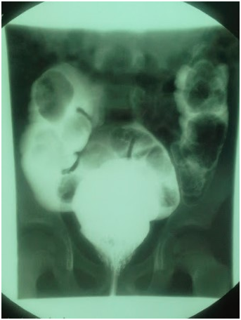
Beside laboratory tests, to investigate more, we noticed his barium enema depicting a narrowed distal colon with proximal dilation (Figures 1-3); retention rectal contrast for longer than 24hours after barium enema also more suggested the diagnosis of Hirschsprung’s disease (HD); although the suction rectal biopsy is considered the current international gold standard for the diagnosis.
He was born symptom-free from natural vaginal delivery of a term non-complicated pregnancy with a birth weight of 3200 g, appropriate for gestational age in a Turkmen family from a consanguineous marriage. There was no other similar problem in other siblings of him in his family. Hirschsprung’s disease is a form of megacolon in a part or the entire intestine due to lack of ganglion cells in myenteric plexus and Meissner plexus.

Figure 3It shows the barium enema depicting a narrowed distal colon with r Hirschsprung’s disease (HD).
The incidence of sporadic Hirschsprung’s disease is 1 in 5000 live births.
In approximately 20% of cases, the diagnosis of Hirschsprung’s disease is made beyond the newborn period. These children have severe constipation, which has usually been treated with laxatives and enemas. Abdominal distention and failure to thrive may also be present at diagnosis.1
Imaging findings of HD after barium enema administration include:2
Presence of transition zone, irregular contractions, mucosal irregularity, and delayed evacuation of contrast material and question mark-shaped colon in total colonic aganglionosis. The differential diagnosis includes acute/chronic megacolon, constipation, intestinal motility disorders, toxic megacolon, etc. Primary supportive measures were prescribed for the patient and he was referred to gastroenterology clinic for further measurement.
None.
Author declares that there are no conflicts of interest.

©2019 Hosseininejad, et al. This is an open access article distributed under the terms of the, which permits unrestricted use, distribution, and build upon your work non-commercially.