MOJ
eISSN: 2379-6162


Case Report Volume 11 Issue 2
1Adjunct Specialist in Oral and Maxillofacial Surgery from Plastic Surgery and Maxillofacial Department at Niño Jesús Pediatric Hospital,Venezuela
2Adjunct Specialist in Oral Surgery from the Burn Center, Plastic Surgery and Maxillofacial Department at Niño Jesús Pediatric Hospital,Venezuela
3Chief Director Specialist in Pediatric Surgery from the Burn Center, Plastic Surgery and Maxillofacial Department at Niño Jesús Pediatric Hospital,Venezuela
4Oral and maxillofacial pathologist. Full professor at dental school, Zulia University,Venezuela
Correspondence: Néstor A Heredia M, Oral and Maxillofacial Surgery from Burn Center. Plastic Surgery and Maxillofacial Department at Niño Jesús Pediatric Hospital, San Felipe,Yaracuy, Venezuela, Tel +58 414 074 7969
Received: June 15, 2023 | Published: July 7, 2023
Citation: Heredia NAM, Ramírez LA, Gutierrez M, et al. Congenital heterotopic gastrointestinal duplication cyst of the face: a rare case report. MOJ Surg. 2023;11(2):93-95. DOI: 10.15406/mojs.2023.11.00230
Gastrointestinal duplication (GDC) cysts have various synonyms; choristoma, enterocystoma, and duplication cyst. They are infrequent congenital benign lesions diagnosed mainly in children in the first decade of life, comprising approximately less than 5% of the pediatric population with a predilection for the male sex. It is exceedingly rare in the oral cavity but it is possible, particularly on the tongue, floor of the mouth, and pharynx. From 1895 until 1994, 30 cases of this lesion in the oral cavity were reported. Nowadays, none of the authors reported a GDC in the facial region. We report an exceptional case of heterotopic gastrointestinal duplication cysts (HGDC) in the facial region of a 13 -years -old male teenager who was referred to our medical department for an evaluation of a painless progressive facial asymmetry. In the first instance, the ultrasound facial soft tissue was made with a 6.5 MHz convex transducer, reporting a big anechoic image with regular borders to a 60 x 26 x 56 mm diameter with a 46cc approximate volume. Following this, a non-contrast magnetic resonance image was executed with an open Philips PanoramaⓇ machine performed at 3T, reporting a hyperintense image in T2-weighted (repetition time msec/echo time msec: 5,500/112) sagittal sequence and FLAIR-weighted (5,714/80) axial sequence; and an isointense image in T1-weighted (24/80) coronal sequence to 54 x 30 x 51 mm diameter compatible with space occupant lesion with interior walls. A complete tumor resection was achieved under balanced general anesthesia, and the specimen was sent for histological examination. Microscopic examination showed a cystic cavity lined mostly by mucous secretory gastrointestinal epithelium and other areas by stratified squamous epithelium. The cystic wall was composed of fibrous connective tissue and contains abundant gastric cells. Its deepest portion exhibits a smooth muscle layer. In summary, HGDC is an unusual congenital head and neck lesion, but it should be considered in the differential diagnosis of neonates and children. The diagnosis of gastrointestinal tract duplications may be suggested by imaging studies; however, the correct diagnosis is defined by histopathologic analysis. Due to the risk of destruction potential and malignant transformation, HGDC should be treated surgically by complete excision.
Keywords: congenital, heterotopic, gastrointestinal, duplication cyst, fac pediatric patient
HGDC, heterotopic gastrointestinal duplication cysts; GC, gastrointestinal cysts; MRI, magnetic resonance image; US, ultrasound
Gastrointestinal duplication cysts (GDC) are rare benign lesion diagnosed mainly in children.1,2 The exact developmental etiology is not clear, but those cysts are thought to arise from displaced embryonic rests of the primitive foregut.3 It is possible that ectopic islands of endodermal cells infiltrate the primitive stomodeum during the fourth week of embryonic development and cause multidirectional differentiation.3,4 It can appear anywhere in the gastrointestinal tract, but they are most commonly observed in the abdomen and thorax. A different site location is named Heterotopic. They are exceedingly rare in the oral cavity, but it is possible, particularly on the tongue, floor of the mouth, and pharynx. Occasionally, they cause airway obstruction or feeding difficulty.2,4,5 From 1895 until 1994, 30 cases of this lesion in the oral cavity were reported. Nowadays, none of the authors reported an GDC in the facial region.1-5 On histological examination, the cyst shows heterotopic islands of gastric or intestinal mucosa covered by columnar ciliated epithelium with a fluid or solid content.2,4 A complete surgical excision is the treatment chosen, and relapses are not expected.4–6 Here, we report an exceptional case of heterotopic gastrointestinal duplication cysts (HGDC) in the facial region of a male teenager.
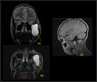
A 13-years-old male was referred to our medical department for an evaluation of progressive facial asymmetry. According to our clinical examination, the asymmetry was caused by a painless tumor on the lateral left face which was extended from the zygomatic region until buccal region and gradually increased in size since the patient was 6-years-old. The lesion was projected into the mouth; the skin and mucosa were normochromic, hard, well defined, adherent to the deep plane and moved on palpation (Figure 1). Open mouth limitation, difficulty during feeding, dysphagia, or odynophagia were not found. In the first instance, ultrasound (US) facial soft tissue was made with a 6.5 MHz convex transducer, reporting a big anechoic image with regular borders to 60 x 26 x 56 mm diameter with a 46cc approximate volume in the left zygomatic arch region. Following this, a non-contrast magnetic resonance image (MRI) was executed with open Philips PanoramaⓇ machine performed at 3T, reporting a hyperintense image in T2-weighted (repetition time msec/echo time msec: 5,500/112) sagittal sequence and FLAIR-weighted (5,714/80) axial sequence; and isointense image in T1-weighted (24/80) coronal sequence to 54 x 30 x 51 mm diameter compatible with space occupant lesion with interior walls; lesion located between maxillary sinus and zygomatic arch bone left, from zygomatic region until buccal region; over to masseter and pterygoids muscles and below to temporal muscle with remodeling bone effects without infiltration (Figure 2); suggesting expansive cyst lesion and recommending histological examination.

Figure 1 Clinical photography showing the facial asymmetry caused by a painless tumor on the lateral left face, extended from the zygomatic region until the buccal region.

Figure 2 (a-b) T2-weighted coronal and axial (repetition time msec/echo time msec: 5,500/112). (c) T1-weighted (24/80) sagittal sequence; showing images compatible with space occupant lesion with interior walls. Yellow arrows point to a lesion located between the maxillary sinus and zygomatic arch bone left, from the zygomatic region until the buccal region, over to masseter and pterygoids muscles and below to the temporal muscle.
A Complete tumor resection was achieved under balanced general anesthesia. Oral intubation was performed with contralateral endotracheal tube location. The intraluminal cyst was aspirated partially and revealed a thick brown/yellow liquid content. A Transoral blunt excision was made and during surgical resection, the mass was identified under the risorius muscle and over the masseter muscle with extension to the depth of the zygomatic region. The lesion did not adhere to adjacent tissues and was fully resected. Haemostasis was achieved using electrocautery, the raw area was covered by mucosal flaps, tissue synthesis was made using polyglactin 910 and the specimen was sent for histological examination.
Macroscopic examination reported a 2,5x1,5x1 cm, opened nodular, dark brown, soft tissue structure, cystic without content. Microscopic examination showed a cystic cavity lined mostly by mucous secretory gastrointestinal epithelium and other areas by stratified squamous epithelium. The cystic wall was composed of fibrous connective tissue and contains abundant gastric cells. Its deepest portion exhibited a smooth muscle layer (Figure 3).
Gastrointestinal duplication (GDC) cysts have various synonyms; choristoma, enterocystoma and duplication cyst. They are infrequent congenital benign lesions diagnosed mainly in children, comprising approximately less than 5% of the pediatric population, most commonly diagnosed in the first decade of life, with a predilection for the male sex.. It agrees with our report of a 13-years-old male patient. Rarely, it can be found in the head and neck; from 57 to 90 reported cases are in the oral cavity.7,8 This difference between ranges reported is a result of the varied nomenclature used for GDC.6–8
Duplication cyst can occur anywhere in the gastrointestinal tract, from the oral cavity to rectum. Commonly subdivided into three categories based on embryological development: Foregut, midgut, and hindgut are more commonly found in the thorax followed by the abdomen.1,3,7,8 The foregut extends from the oral cavity to the proximal duodenum up to the attachment of the bile duct and also includes the liver, gallbladder, and pancreas. These are subsequently categorized into esophageal, gastric, bronchogenic, and neurenteric cysts based on their embryological origin.8 Gastric duplication cysts (GsDC) comprise about 4–9% of all enteric duplication cysts. Approximately 67 % of GsDC are identified within the first year of life, are generally asymptomatic and are encountered as incidental findings. Most GsDC are related to the greater curvature of the stomach, as duplications are more common dorsal to the primitive gut and their mechanism of formation, it is not well understood. These are usually solitary, they do not communicate with the gastric lumen and are considered to be a result of abnormal canalization.8,9 An unusual location of the tumor on the lateral left face, extended from the zygomatic region until the buccal region had not been reported until now, but the varied nomenclature used for GDC, which has a fail to account for all reported cases. We think that it is probably a tumor that developed in the mouth and grew towards the face. Clinically, patients are usually asymptomatic, but they can sometimes develop secondary symptoms with a mass effect on adjacent structures, including non-productive cough, stridor, chest pain, or dysphagia.3,4,6,8 In agreement with the findyings, our patient didn't had symptoms, only showed an facial asymmetry caused by a painless tumor.
Ultrasound is an active diagnostic and therapeutic aid available for head and neck pathology. Because computed tomography exposes children to ionizing radiation, and MRI on occasions requires sedation, US has become the favored modality by many clinicians. It does not only obviate radiation exposure and the need for sedation, it is less expensive and easier to execute than the other two imaging options. Proponents of US in this clinical setting also point out its utility in simultaneously evaluating ectopic tissue. 10,11 An uncomplicated cyst is usually anechoic on US, with internal echoes potentially indicating hemorrhage or pus. A classic pattern of alternating hyperechoic and hypoechoic layers is frequently seen on ultrasonography (sometimes referred to as the“gut signature sign”).
MRI findings include a low-intensity cyst wall due to its fibrous nature, low to intermediate intensity on T1-weighted imaging, and high signal intensity on T2-weighted imaging. As a particular image, we describe an isointense image in T1-weighted (24/80) coronal sequence compatible with space occupant lesion within intracystic signal intensity interior walls, with remodeling bone effects without infiltration. These MRI characteristics could be related to the progressive growth of the clinic referred by our patient. In future studies it will be important to correlate.
On histopathology, the esophageal duplication cysts are lined by squamous epithelium and have a double layer of smooth muscle in their wall.7,8 The GsDC shows a cyst wall composed of mucosa and submucosa, predominantly gastric type and mucus cells. We found a cyst lined by squamous epithelium, which contains abundant gastric cells, and its deepest portion exhibits a smooth muscle layer; it appears to be a common feature between them. Though typically benign, some authors reported histologic findings of malignant transformation.7–9 It doesn’t happen in our case.
Complete cyst excision is the treatment of choice in symptomatic cases. The role of surgery in asymptomatic cases is less clearly defined.7,8 However, a progressively expanding lesion that causes facial asymmetry should be considered a surgical indication.
In summary, HGDC is an unusual congenital head and neck lesion, but it should be considered in the differential diagnosis of neonates and children. Can appear anywhere from the gastrointestinal tract, be extended from the oral cavity to the face, and are kindred with the foregut. The diagnosis of gastrointestinal tract duplications may be suggested by imaging studies. However, the correct diagnosis is defined by histopathologic analysis. Due to the risk of progressive growth, destruction potential, and malignant transformation, HGDC should be treated surgically by complete excision.
Chief's Director’s from Niño Jesús Pediatric Hospital; Medical Residents from the Burn Center, Plastic Surgery, and Maxillofacial Department at Niño Jesús Pediatric Hospital and all those people made possible this research.
The authors declare no conflicts of interest.
This research complies with the World Medical Association Declaration of Helsinki on medical protocols and ethics. As the images of the patient were essential to this paper, the patient’s mother gave written consent.

©2023 Heredia, et al. This is an open access article distributed under the terms of the, which permits unrestricted use, distribution, and build upon your work non-commercially.