MOJ
eISSN: 2379-6162


Case Series Volume 11 Issue 2
Department of Surgery, School of Medical Sciences, Universiti Sains, Malaysia
Correspondence: Mohamed Shafi Mahboob Ali, Department of Surgery, School of Medical Sciences, Universiti Sains, Malaysia, Tel +601114724649
Received: May 16, 2023 | Published: June 8, 2023
Citation: Ali MSM. Clinicopathological characteristics in male breast cancer: a case series and literature review. MOJ Surg. 2023;11(2):76-80. DOI: 10.15406/mojs.2023.11.00227
Male breast cancer (MBC) is a rare entity and overall cases reported were less than 1%.However, the incidence of MBC is rising regularly every year. Due to the lack of data on MBC, the diagnosis and treatment are tailored similar to female breast cancer. MBC risk increases with age and is usually diagnosed 10 years late as the disease progression is slow compared to female breast cancer (FBC).Commonest features of MBC is intra-ductal variant and often, upon diagnosis, the stage of the disease is already advanced. Prognosis of MBC is often poor but with the recent knowledge and advancement, new modalities of treatment are emerging.
Keywords: male breast cancer, clinicopathology
Breast tissue is underdeveloped in males compared to females. In an adult male, the breast tissue usually composed of fat tissues, ducts and no lobules.1 It has been reported that after puberty there is about 10% of male adults has persistent breast tissues.2 Early detection of MBC is difficult as the disease usually presents late in the lifetime. In a population-based studies done by Anderson et al, the mean age of MBC diagnosis was 65.8 years old.3 Due to its rarity and lack of data, it is difficult to understand the character and behavior of MBC. Hitherto, we are referring to female breast cancer guidelines in navigating MBC management. The risk for MBC increases with advancing age and the prognosis is worse compared to female breast cancer based on overall survival.4,5 MBC is closely related to BRCA2 mutation with 6.3% of lifetime absolute risk for breast cancer.6 Several studies shown that most of the MBC are of a ductal variant with ER-positive and Luminal type A.7 We would like to report the clinicopathological characteristics of five male breast cancer cases that we encountered in our centre.
A 72 years old gentleman presented to our centre in 2019 with the right breast swelling and painless nipple bloody discharge for the past three months. He claimed the swelling was progressively increasing in size with changes noted on the surface of the nipple skin. The patient also had constitutional symptoms such as loss of weight and loss of appetite for the past one year. He claimed he lost about 10kgs in the last 6 months. Otherwise, the patient denies having any family history of breast cancer nor consumed any kinds of hormonal pills or supplements. Triple assessment was done on the patient and clinical examination revealed a hard, non-tender lump at the retro areolar region measuring about (2x1) cm. There were blood and serous discharge from the nipple. No mass was palpable on the other region of the breast or contralateral side. Bilateral axillary lymph nodes were not palpable. Initial right breast ultrasound noted the presence of cystic and solid lump at the retro areolar region measuring (1x1x1.8) cm. Also noted was internal vascularity with a layer of sediment within the cystic mass. The imaging findings were categorized as BIRADS 4.We then proceed with computed tomography (CT) scan and noted the presence of a focal enhancing retro areolar lesion on the right breast measuring (0.8x0.8x1.0) cm with no extension to the underlying structures. There were some sub-centimetres bilateral axillary lymph nodes with preserved fatty hilum with largest was 0.9cm in the right axilla. Ultrasound-guided biopsy was performed and was confirmed to be of intra-ductal papillary carcinoma in origin. The patient was staged as T1N0M1 according to the TNM classification. The patient was arranged for a right mastectomy the following week. He was then discharged home well after a week and a surveillance mammogram was performed a year later and no focal lesion seen on the left breast (Figure 1 & 2).
A 59 years old gentleman who was an ex-chronic smoker (40 pack-years) was referred to our centre for a huge left breast mass. On further history, the patient claimed that the left breast swelling has been there for the past one year and he never seek any medical treatment. While in the ward patient developed generalized tonic-clonic seizure (GTC) for six episodes, aborted by intravenous Phenytoin. An urgent brain computed tomography (CT) was performed and a left frontal intra-axial space-occupying lesion was found. Clinically, the left breast swelling was irregular in shape, measuring about (18x5) cm, hard, non-tender and fixed to the underlying structures. There were left axillary lymph nodes palpable. A TRU-CUT biopsy of the left breast mass was performed and histopathology results came out as Invasive breast carcinoma of no special type (NST).Contrasted tomography (CECT) of the thorax was performed for the staging purpose and was reported as the presence of lobulated heterogeneously enhanced mass occupying the whole left breast measuring (11.1x4.7x9.2) cm with an erosion of the left anterior end of second to fourth lateral surfaces of the ribs. Also, there were multiple enhancing axillary lymph nodes bilaterally, at the pre-tracheal, para-tracheal, sub-carina and the presence of lung nodule in the superior segment. There was metastatic lesion seen on the left adrenal gland measuring (3.7x2.9x3.5) cm as well. The patient was staged as T4N1M1. No surgery was done as patient deteriorated and succumbed to death due to Pulmonary Embolism (PE) on day eleventh of admission (Figure 3–5).
A 74 years old gentleman with underlying hypertension and dyslipidemia presented to our Centre with a right breast swelling for the past 2 years. The patient claimed that the swelling been progressively increasing in size associated with bloody nipple discharge and foul smell. He never seeks any treatment since then. Clinically there was a (4x4) cm fungating mass at the right nipple-areolar complex (NAC), hard, non-tender and fixed to the underlying structures. The lesion demonstrated contact bleeding during manipulation. Mobile right axillary lymph nodes were palpable. A wedge biopsy was performed the next day and the histopathology result came out to be Invasive breast carcinoma, no special type (NST).Computed tomography (CT) of the thorax, abdomen and pelvis was arranged for the patient. It was reported as right breast mass with local extension to the underlying pectoralis major muscle with right axillary lymphadenopathy. Also reported was bilateral lung nodules which suggestive of metastasis (T4CN1M1). A right mastectomy with axillary dissection was performed, a total of 15 axillary lymph nodes were removed and sent for histological examination. Microscopic examination of the specimen was described as ductal carcinoma in-situ (DCIS) of high-grade nuclear feature comprising of cribriform and comedo-necrosis pattern. There were five out of seven lymph nodes positive for tumour infiltration. The patient was subsequently referred to the oncologist and a regime of six cycles of FEC (5-Fluorouracil, Epidoxorubicin, Cyclophosphamide) and 15 fractions of radiotherapy was planned (Figure 6, 7).
A 64 years old gentleman with an underlying Stanford Type B dissecting aneurysm post endovascular intervention (EVAR) was referred to our breast clinic for a right breast lump. According to the patient, the lump has been there for the past three months and it is gradually growing bigger .The right breast lump was associated with pain and he was dependent on regular pain killer. Otherwise, the patient denies any history of trauma, discharge per nipple or a contralateral breast lump. There was no positive family history of breast cancer as well. Upon clinical examination, there was a round swelling at nine o’clock position, 1 cm away from the right nipple, measuring about (3x3) cm in size. The mass was firm, mobile and non-tender. There was no nipple discharge or Peau de’orange skin changes seen. Right breast ultrasound showed the presence of a well-defined lobulated heterogeneous mass at 9 o’clock, 2 cm from the nipple. It measures (1.9x3.2x2.7) cm with posterior enhancement and microlobulation. There was internal vascularity with the resistive index of 0.81(BIRADS 4).Otherwise no dilated ducts are seen and no enlarged right axillary lymph nodes either. The left breast showed no focal lesion. An ultrasound-guided biopsy was performed on the same day and the histopathology result revealed the lesion as Invasive breast carcinoma, no otherwise specified (NOS).The patient was counselled to undergo a computed tomography (CT) scan as the ultrasound findings were suspicious. The CT scan was reported as right breast invasive ductal carcinoma with evidence of local infiltration and no distant metastasis (T4N0M0). The patient was offered a right mastectomy but unfortunately patient defaulted our follow-up (Figure 8, 9).
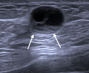
A 67 years old male with a previous history of right nipple discharge and underwent right microdocechtomy in 2012 presented to our Centre in late 2020 with a right breast lump. According to the patient, he started to notice the lump three months ago. The lump does not grow in size, otherwise, he experiences some occasional pain. He also denies any bloody nipple discharge, erythema or contralateral breast lumps. Initial right breast ultrasound demonstrated a well-defined rounded, anechoic lesion with posterior enhancement at 9 o’clock, periareolar region measuring (1.1x1.4x1.4) cm in size. There were internal septations with an irregular solid component within the cyst. Otherwise, there is no intralesional vascularity or calcifications seen. An ultrasound-guided biopsy of the lesion done and the result was suggestive of intraductal papillary carcinoma (DCIS).TNM staging for the patient was T1bN0M0. The patient underwent a right mastectomy and sentinel lymph node dissection. The sentinel lymph node was identified using a combination of technetium 99 tag and methylene blue dye. The dissected sentinel lymph node was sent for frozen section and it came out as negative for malignancy, thus no axillary clearance was done. Histopathology report revealed that all resection margins were free from tumour and no evidence of metastasis in the axillary lymph node sent. The patient was started on Tamoxifen and planned for surveillance computed tomography (CT) Thorax, Abdomen and Pelvis (TAP) (Figure 10) (Table 1).

Figure 10 Ultrasound image showing a well-defined rounded, anechoic lesion with posterior enhancement at right breast. Presence of solid component (white arrows) noted within the cyst.
|
Features |
A |
B |
C |
D |
E |
|
Age(years) |
72 |
59 |
74 |
64 |
67 |
|
Histopathology |
Intraductal Papillary carcinoma (dcis) |
Invasive carcinoma of no special type (nst) |
Invasive carcinoma of no special type (nst) |
Invasive carcinoma no otherwise specified (nos) |
Papillary carcinoma of no special type (nst) |
|
Multifocality |
No |
No |
No |
No |
No |
|
Bilaterality |
No |
No |
No |
No |
No |
|
Side |
Right |
Left |
Right |
Right |
Right |
|
Type of surgery |
Right mastectomy |
No surgery done |
Right mastectomy and axillary clearance |
No surgery done |
Right mastectomy and sentinel lymph node dissection |
|
Axillary lymph Node dissection |
No |
No |
Yes |
No |
Yes |
|
No. Of positive Lymph nodes |
Nil |
Nil |
5 out of 7 |
Nil |
Nil |
|
Stage |
T1N0M1 |
T4N1M1 |
T4CN1M1 |
T4N0M0 |
T1bN0M0 |
|
Tumor grade |
Grade 2 |
Grade 3 |
Grade 3 |
Grade 1 |
Grade 2 |
|
Lymphovascular Invasion |
No |
No |
Yes |
No |
No |
|
Hormone receptor status |
ER: positive PR: positive E-cadherin: Positive |
ER: Negative PR: Negative |
ER: Positive PR: Positive |
ER: Positive PR: Positive |
ER: Positive PR: Positive |
|
Her-2 status |
Negative |
Negative |
Positive |
Positive |
Negative |
Table 1 Clinicopathological features
Male breast cancer (MBC) is a rare type of neoplasm with overall cases of less than 1%.However, an annual increment of MBC has been reported in the U.K, the U.S and Europe.8 There were no large scale multicenter studies conducted in South East Asia for MBC probably due to its rarity in this region, thus, we are lack of the necessary data. MBC pathology closely resembles the female breast cancer. The mean age of MBC in our case series is 67.2 years old. MBC is diagnosed later in male compared to females and the prognosis upon diagnosis is usually poor. There are several risk factors related to the MBC, such as high estrogen level, radiation exposure, strong family history of breast cancer and some rare genetic disorder such as Klinefelter’s syndrome. Undescended testis, orchidectomy, orchitis and late puberty has been related to MBC as well.9 In our case series, most patients were diagnosed with ductular invasive breast carcinoma of no special type(NST). All patients have no multifocality nor bilaterality disease. Unfortunately, most of the patients presented to our Centre at the late stage of the disease, some, with distant metastasis. Another important clinicopathological aspect that we looked upon was the tumour grade. There are several techniques for determining a tumour grade. Frequently used is Bloom-Richardson grading system which takes into account the gland feature, nuclear features and mitotic activities. Most of our patients in this case series have grade 2 and 3, in other words, has high mitotic activities and the tumor is actively proliferating. Out of five patients, one patient had a mastectomy while another two patients had mastectomy and axillary dissection. Patient B was not operated as he succumbed due to pulmonary embolism (PE).While patient D defaulted his follow-up since last year and was not contactable. Only patient C has evidence of lymphovascular invasion out of five patients. Computed tomography (CT) scan of patient C demonstrated that he had bilateral lung metastasis. Patient B has been detected to have a brain metastasis along with breast tumour despite having no evidence of lymphovascular invasion. Brain metastasis of patient B in this case series was diagnosed less than six months and the tumour can be labelled as a synchronous tumor. Synchronous tumor in a breast cancer patient has been reported in several case reports.10 Most of our MBC patients’ demonstrated receptors positive for both Estrogen and Progesterone except patient B. This correlates with most studies where MBC has a high ER/PR positivity(Dimitrov, Colucci and Nagpal, 2007).Only patient C and D showed positive for human epidermal growth factor receptor 2 (HER-2) while the rest were negative. Both patients (C and D) were T4 stage and ER/PR positive. Patient C underwent surgery and adjuvant therapy while patient D defaulted follow-up. Targeted therapy using monoclonal antibodies such as Herceptin has shown a promising result in HER-2 positive MBC patients.11 Patient B is the only patient with triple negative receptor status in our case series. In triple-negative breast cancer (TNBC) the cancer cells do not demonstrate estrogen or progesterone receptors nor produce enough HER-2 proteins. Treating a TNBC could be a great challenge especially in the case of male breast cancer with a very limited data available. New studies have emerged on Novel-ADC (antibodies drug conjugate) in treating TNBC which can be applied on aggressive MBC with triple negative.12
Male breast cancer (MBC) is as fatal as female breast cancer (FBC).The scarcity of cases, lack of data and the long period of disease progression making it complicated for the clinicians to manage. In our case series, most male breast cancer cases appeared at the late stage of life with an advanced tumour grade, often, with distant metastasis and poor prognosis. What is promising, the majority of these patients demonstrated Estrogen and Progesterone receptors positivity which may respond to hormonal treatment with an acceptable outcome. Other techniques for studying the disease using immunohistochemical and tumour genetics should be explored. Larger multicentres local and international collaborations needed in gathering more data and information about male breast cancer (MBC).Local health facilities and the governing bodies should play a vital role in increasing the awareness of male breast cancer as well as conducting a screening programme in the community as MBC is a treatable and preventable disease.
None.
The authors declare no conflicts of interest.

©2023 Ali. This is an open access article distributed under the terms of the, which permits unrestricted use, distribution, and build upon your work non-commercially.